Absorption in the digestive tract.
Absorption is the process of transition of substances from the gastrointestinal tract into the blood and lymph through cells, their membranes and intercellular passages.
Occurs throughout the gastrointestinal tract, but in different parts of it with varying intensity.
The mucous membrane of the oral cavity is capable of absorbing, but there are usually no end products of the breakdown of nutrients in the oral cavity. Some medicinal substances are well absorbed here.
Water, mineral salts, monosugar, medicinal substances, alcohol, very few amino acids are absorbed in the stomach.
The main absorption process takes place in the small intestine.
Carbohydrates absorbed into the blood as glucose and other monosaccharides.
Squirrels enter the bloodstream in the form of amino acids. Neutral fats are broken down by enzymes to glycerol and fatty acids. Glycerin is soluble in water, therefore it is easily absorbed. Fatty acids are absorbed only after interaction with bile acids, with which they form complex compounds. Fats go mainly to the lymph and only 30% to the blood.
The large intestine absorbs water and mineral salts.
Suction mechanisms.
Passive transport (diffusion, filtration).
Active transport, with the participation of carrier enzymes.
Chewing- is done reflexively. Food in the mouth irritates the receptors, from them signals are transmitted along the afferent fibers of the trigeminal nerve to the center of chewing (medulla oblongata). As a result, the food is crushed, in addition, it is mixed with saliva and a food lump is formed.
Swallowing- a reflex act, its center is in the medulla oblongata. In the process of swallowing, there are 3 phases:
1. Oral (arbitrary). The food lump moves to the back of the tongue with movements of the tongue and cheeks, then successive contractions of the muscles of the tongue of the anterior, middle and posterior groups move it to the root of the tongue.
2. Pharyngeal (fast involuntary. Irritation of the receptors of the mucous membrane of the root of the tongue reflexively causes contraction of the muscles that lift the soft palate, the muscles of the tongue and muscles that lift the larynx. In the oral cavity pressure increases, so food moves into the pharynx. Then the muscles of the pharynx begin to contract above the food lump and it moves to the esophagus , pressure in the pharynx increases, the pharyngeal-esophageal sphincter opens and food passes into the esophagus.
3. Esophageal (slow involuntary). The passage of food through the esophagus occurs due to successive contractions of the annular muscles in the wall of the esophagus. They have the character of a wave that occurs in the upper part of the esophagus and spreads towards the stomach. This type of contraction is called peristaltic. The regulation of motility is carried out by the autonomic nervous system: the parasympathetic vagus nerve enhances the peristalsis of the esophagus and relaxes the cardiac sphincter at the border with the stomach, the sympathetic nerves inhibit peristalsis and increase the tone of the cardiac sphincter.
Motor function of the stomach.
Provided by the work of smooth muscles. There are 3 types of physical activity in the stomach:
1. Peristaltic movements occur due to contractions of the circular muscles. The wave of contraction begins in the area of the cardiac part of the stomach and goes to the pyloric sphincter. Wave frequency -3 times in 1 min.
2. Systolic contractions are muscle contractions in the pyloric region of the stomach. Provide the transition of the chyme into the duodenum.
3. Tonic contractions are caused by changes in muscle tone in different parts of the stomach. As a result, the food mass is mixed with the digestive juice and moves to the exit from the stomach.
The parasympathetic nervous system enhances motor skills, the sympathetic nervous system inhibits. Humoral factors that enhance motor skills: insulin, gastrin, histamine. Humoral factors that inhibit gastric motility: enterogastrin, cholecystokinin, adrenaline, norepinephrine.
In addition to the named types of contractions in the stomach, there are antiperistalsis, which occurs with vomiting.
Transfer of food from the stomach to the intestines.
Food is in the stomach for 6 to 10 hours. During this time, the smooth muscles in the wall of the stomach contract, the contents of the stomach are mixed with gastric juice, moves towards the exit into the small intestine and exits into the duodenum.
Chyme enters the duodenum in portions from the pyloric stomach. There is a sphincter on the border between the stomach and the duodenum. Hydrochloric acid of gastric juice irritates the receptors of the gastric mucosa in the pyloric section, the sphincter opens, the muscles in the wall of the pyloric section contract and the chyme passes into the duodenum. Here, the reaction of the medium is weakly alkaline, therefore, the acid in the chyme acts on the mucous membrane of the duodenum, the sphincter contracts and the evacuation of the chyme from the stomach to the intestine stops. When the reaction of the environment in the intestine is restored, the process is repeated.
The motor function of the digestive tract. It is with this function that the process of food absorption, chewing, swallowing, movement of food contents along the digestive tract is associated. This function facilitates mixing of food with digestive secretions. It is necessary for absorption and for the removal of indigestible residues. Various methodological approaches are used to study the model of the digestive tract.
Balloon kinetography. The introduction of a balloon into the alimentary canal, connected to a monometer using a system of tubes. In humans, the X-ray method of investigation with the preliminary introduction of barium sulfate is widespread.
The method of electrogastrography is used, based on the registration of electrical impulses. The experiment uses contractions of isolated areas of the digestive tract, visual observation.
A person also uses the auscultation method - listening to sounds associated with motor skills.
In children, the act of sucking is also referred to as motor function. After placing food in the mouth, chewing begins. Chewing consists of a reflex movement of the lower jaw in relation to the upper jaw. The chewing muscles include: the chewing muscles proper, the digastric, temporal, superior and inferior pterygopalatine.
When the mouth is opened, the proprioceptors of the masticatory muscles are irritated and, at the same time, a contraction of the masticatory muscle proper, and the temporal, pterygopalatine, occurs reflexively.
If food is in the oral cavity, then it irritates the receptors of the mucous membrane, then this causes contraction of the digastric muscle, which contributes to the lowering of the lower jaw. In addition, it also descends due to gravity.
The chewing function makes it easier to swallow food, destroys the cellulose membrane of fruits and vegetables, increases the area of contact with digestive enzymes, promotes mixing and wetting of food with saliva, and creates better contact with taste buds. Chewing helps to release food odors. The smell acts on the olfactory receptors, and this increases the pleasure of eating.
As a result of chewing, a food lump is formed, which is swallowed.
There are 600 swallowing acts per day. 200 when eating, 350 the rest of the time, 50 at night.
The act of swallowing is divided into an arbitrary phase (before the food moves to the root of the tongue). When the lump of food passes behind the root of the tongue, the involuntary phase of the act of swallowing begins. Food irritates the sensory receptors in the mouth formed by the trigeminal nerve. The taste buds, which are associated with the 7th pair, and the back third with the 9th pair. The vagus also takes part in the sensitive innervation. From these receptors, sensory impulses go to the center of swallowing. And already from there, along the motor fibers of the same nerves, a coordinated muscle contraction occurs, in which the soft palate rises and closes under the nasopharynx. The trachea and hyoid bone rise, the epiglottis descends and this closes the airway. The root of the tongue rises, presses against the palate and makes it impossible for the food lump to return to the oral cavity.
The pharyngeal phase of swallowing begins. Contractions of the pharynx move the lump towards the esophagus. At the border of the pharynx and esophagus is the upper esophageal sphincter. It occupies a segment with a length of 3 centimeters. With the contraction of the pharyngeal muscles, the upper esophageal sphincter opens. Thus, the food bolus enters the esophagus, through which the next, esophageal phase of the act of swallowing is already taking place. The movement of the food lump along the esophagus is associated with the muscles of the esophagus. In the upper third, this will be the striated muscle. And the lower ones are smooth. Distinguish between circular and longitudinal muscles.
The speed of movement of the food lump is 4-5 cm per second. Solid food passes the esophagus in 8-9 seconds. In this case, a high pressure is created inside the esophagus (from 30 to 120 mm).
If a person consumes liquid food, then the tone of the muscles of the esophagus decreases and a lumen is created through which a column of liquid enters. This process takes 1-2 seconds.
When the esophagus passes into the stomach, there is a cardiac sphincter. He is in a state of tonic tension. The sphincter tone is maintained due to nervous and hormonal influences (gastrin, cholicitokenini, matenin). The pressure created by the sphincter is 10-15 mm. As the food bolus approaches the sphincter, it relaxes. This makes it possible for the food bolus to pass into the stomach. Simultaneously with the relaxation of the cardiac sphincter, there is a relaxation of the tone of the stomach muscles. Receptive relaxation. The muscles of the esophagus are innervated by the vagus nerve, which promotes motor skills, but the vagus nerve does not relax the sphincter. With a high tone of the muscles of the esophagus, a state of acolosia can occur, when food is retained in the lower part of the esophagus and causes the expansion of this part.
Reflux is the throwing of stomach contents into the esophagus. This condition is accompanied by a sensation of heartburn. If this happens frequently, ulceration of the esophagus may occur. In case of sphincter insufficiency, a state of aerotopia can be observed - swallowing air with food. This is especially evident in infants during sucking. Therefore, the child should not be immediately placed in a horizontal position after sucking, because this will promote regurgitation.
Stomach motility. The motor function of the stomach is related to the function of smooth muscles. Located in three directions: circular, longitudinal and oblique. The stomach is separated from the esophagus. The exit from the stomach from the duodenum is separated by the pyloric sphincter. The functional prepyloric sphincter is also distinguished. The smooth muscles of the stomach receive innervation from the vagus nerve and the sympathetic nerve. In addition, the stomach has local innervation due to the submucosal and muscleless plexus. In this case, cells of the first type can perform as an exciting function. Stomach motility is represented by tonic contractions of smooth muscles, wave-like peristalsis contractions, and smooth muscles also have the property of automaticity. Separate smooth muscle cells are connected to each other using tight electrical contacts, which allows smooth muscles to function as a functional sensidia. Locomotor activity in the stomach is observed during digestion. But the contraction in the stomach is also observed without food. Such motility is called fasting-periodic motility.
During the first meal, there is a decrease in stomach tone. This will be a receptive relaxation of the stomach muscles, which creates reservoirs for food in the stomach. In this case, each next food lump falls into the center of the previous one, due to which a layered stomach contents occur.
After the act of eating ends, there is a gradual increase in the tone of the stomach muscles. With an increase in the tone of the stomach muscles, peristaltic contractions begin to appear. Motor function is differently expressed in different departments. In the proximal part (includes the bottom and upper third) tonic contractions are better expressed. And the distal part, which includes the lower rub, has a greater ability to wave-like contractions. Stomach motility contributes to the placement of food in the stomach, grinding food inside the stomach, mixing with gastric juice.
The basic rhythm is 3 contractions per minute. Moreover, peristaltic waves can travel at a speed of 0.3 to 4 contractions. At the beginning, peristalsis in the stomach is not deep. More frequent contractions are observed. As the peristaltic wave advances, its strength increases towards the pyloric section. At this stage, mixing and machining take place. As the contractions intensify, the rhythm decreases, and the peristaltic waves become more powerful. Part of the digested food is pushed through the pyloric sphincter into the duodenum. But particles no more than 1 mm in diameter can pass into the duodenum. Entering the intestine causes a powerful contraction of the pyloric sphincter and contraction of the pyloric section. In this case, the content is thrown into the body of the stomach. The return of the sorbent to the body of the stomach is retro-pulsation. With this backward movement, further fragmentation of the particles occurs.
The process of evacuation of food from the stomach will be determined by the coordinated work of the muscles of the stomach and the digestive sphincter. The transition process will be influenced by the volume of gastric contents, the chemical composition and color of food, the consistency, the degree of its acidity and osmotic concentration. In order for the contents of the stomach to pass into the duodenum, it must be liquid or semi-liquid. It should also have isotonic pressure and a certain degree of acidity. When food enters the duodenum, the mucous receptors are irritated. Irritants can be fatty acids, osmotic pressure, etc. With irritation, an obturator reflex occurs, which consists in closing the pyloric sphincter and weakening gastric motility.
The accelerated flow of food from the stomach into the intestines leads to dumping syndrome, which is characterized by the appearance of severe weakness, dizziness, and the desire to lie down after eating.
In a fasting state, periodic contractions appear in the stomach (migrating myoelectric complex). Occurs every 90 minutes and lasts 3-5. The migratory complex manifests itself not only in the stomach, but also in the small intestine. The significance of these contractions is due to the fact that the mucous membrane is freed from mucus, food debris and dead cells. These contractions coincide with the feeling of hunger.
Periodic hunger motility is associated with hunger in the hypothalamus. It is felt by changes in the blood (the level of glucose, calcium decreases, the appearance of choline-like substances).
The impulses are directed to the cerebral cortex. At the same time, there is an influence on the underlying departments.
Small intestine motor function. In the wall of the small intestine there is an external longitudinal and an internal circular. Distinguish between tonic contractions, rhythmic segmentation, pendulum contractions and peristalsis contractions. Rhythmic segmentation is manifested in the rhythmic contractions of the circular muscles. At the same time, it is segmented into separate sections.
Pendulum contractions involve not only circular muscles, but also longitudinal ones. The contraction of the circular muscles causes contraction, and the longitudinal muscles cause expansion.
The frequency of contractions in the upper sections is reduced by 10-12 per minute. And in the lower sections it will be 5-8. Perstalsis is needed to move the contents of the small intestine in the distal direction.
With a slow contraction, the speed is equal. With a rapid peristalsis, the speed reaches 7-21 cm.
Small intestine motility depends on the composition of the food. Coarse foods stimulate motor skills; fatty foods also increase motor skills. Serotonin, histamine, gastrin, methylin, cholicystekinin, substance P, vasopressin and bile stimulate. To inhibit include gastrointestinal and vasointerstinal. The motor function of the small intestine is controlled by the integral part of the autonomic nervous system.
The contents of the small intestine only travel in one direction. Antiperestaltic contractions are observed only during vomiting.
Contractions begin 1-4 minutes after a meal every 30-60 seconds, the sphincter reflexively expands and the contents flow from the small intestine into the blind. The work of this sphincter occurs due to the gastroiliocytic reflex. These two areas are related to each other.
When food enters the large intestine, approximately the same pattern of motor activity is observed in the large intestine as in the small intestine, but the movement takes place much more slowly. In addition, anti-perestaltic contractions are also present here. Therefore, in the course of the motor function, the contents slowly move one way or the other. This promotes the absorption of water, the formation of feces. Small amounts of nutrients are absorbed. Approximately 3-4 times a day, there are propulsive contractions of the colon, which push the contents in the distal direction. Colon motility regulation is carried out by local plexuses, as well as parasympathetic and sympathetic nerves. Formed feces are collected in the distal colon before reaching the rectum.
In humans, the desire for defication occurs when feces enter the rectum. The first sensations arise when the pressure in the rectum rises to 18 mm Hg. There are 2 sphincters in the rectum. Internal (smooth muscle) and external (striated muscle). Both sphincters are in a tonic state. The sphincter tone is controlled by the sacral section of the parasympathetic system. The spinal center is also associated with the overlying centers. But the centers of the brain mainly have an inhibitory effect. The activity of these centers allows for arbitrary regulation of the act of deification. When the mucous membrane is irritated, there is a reflex increase in the activity of the parasympathetic centers, which enhances peristalsis and relaxes the internal sphincter.
The defecation reflex intensifies after eating. The suppression of this reflex can lead to impaired patency. Arbitrary regulation is established at 2 years of age. When the spinal cord is damaged above the sacral region, the deflection reflex occurs periodically, but involuntarily. The defeat of the sacral region leads to relaxation of the sphincter.
Food is a vital necessity for a person. Its usefulness, timely admission in sufficient quantities ensures the normal functioning of the whole organism, emotional state and performance. The functions of the stomach are of prime importance for these purposes.
In order to understand how the stomach works, you need to familiarize yourself with its anatomy, the structure of cell structures, and the muscle layer. Knowledge of physiology helps to find the right approach in the treatment and prevention of certain diseases, not only of the stomach, but of the entire digestive tract.
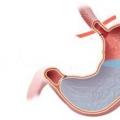
The stomach is a hollow, muscular organ lined with a mucous membrane from the inside with a secretory and enzymatically active layer. It is one of the key organs of the gastrointestinal tract, where deep processing of food with enzymes, gastric juice, digestion of the food lump, from which nutrients are absorbed into the blood take place. Then, with the help of contractile, translational movements - motility, the food lump moves further into the intestine, where the final stage of processing and the formation of feces occurs.

Digestion begins in the mouth, where food is chewed and processed with enzymes. Then, through the esophagus, it enters the stomach cavity, which is conventionally divided into three sections:
- cardiac;
- fundic;
- gatekeeper.
The cardiac region has a sphincter that opens when food enters the vestibule of the stomach. After the lump has penetrated inside, it tightly closes the opening, preventing gastric acid from entering the lower esophagus.
The fundus is the main area of the organ, which is supplied with a secretory layer on the mucous membrane. When food enters, the secretion of hydrochloric acid, gastrokinetics, which stimulate the peristaltic movements of the stomach, is activated.
The gatekeeper or antrum is the final passage of the stomach into the duodenum. Digested food, moving along the stomach cavity, stimulates the opening of the pylorus sphincter for its release into the duodenal lumen.
A very important moment at this stage is the complete closure of the pylorus flaps to prevent the throwing of bile into the stomach cavity. If there is an inferiority or defect in the sphincter due to operations, regular overeating or other reasons, then bile can corrode the walls of the stomach, gradually leading to the development of erosive gastritis, then to an ulcer.
The muscular layer of the stomach is a smooth muscle that does not obey the will of a person, and contractions and movements occur only on the basis of natural mechanisms. That is why it is important to understand the structure of the organ, because you cannot consciously force the stomach to contract if its physiological mechanisms are damaged or lost.
Cells that have enzymatic and secretory activity are also susceptible to damaging effects. Inadequate production of enzymes due to external influences, internal causes, age-related changes leads to insufficiency of the functions of the human stomach.
Digestive functions
It is clear that the main task of the stomach is to digest food and move it further. But this is a too general concept, such an approach does not allow correctly diagnosing, treating and developing measures for the prevention of his diseases. The stomach has the following digestive functions:

Each of them is necessary for full-fledged digestion, providing the body with vitamins and building materials. Good digestion, absorption and promotion of food is especially important for newborn children, whose body is just starting to work, therefore, baby nutrition and health should be given the utmost attention.
During pregnancy, taste preferences change, there is a complete restructuring of all organs and systems, therefore, the lack of any of the functions can affect the health of the unborn baby or mother.
Depositing
Translation from Latin means "accumulation", that is, food is retained in the stomach for a while. This is necessary so that all nutrients are properly processed, the blood rushes to the walls of the organ, and the process of digesting food goes as expected. If there was no mechanism for the delay of the food lump inside the stomach for several hours, then it would fall further, without mixing with the enzymes, hydrochloric acid contained in the gastric juice.
The depositing function of the human stomach is provided due to the mechanism of reflex relaxation of the muscular apparatus of the fundus. The retention of chyme (food lump) is carried out for a sufficiently long time: from 3 to 10 hours, depending on the density of the food received.
Motor
This is a whole range of types of motor mechanisms, due to which the entire volume of food that has entered the stomach is digested and gradually moves further. The work of the stomach at this moment is performed due to peristaltic waves, topical contractions of the fundus and body of the stomach, systolic contractions of the pyloric section.
 During the time of movement, food components continue to dissolve, be digested and processed by gastric juice. The result of this functional work is the complete dissolution of food components.
Suction
This is one of the most important tasks: nutrients necessary for humans are extracted from food products and they must enter the bloodstream, so that due to their delivery to the target organs, the corresponding metabolic processes take place:
- proteinaceous;
- fatty;
- carbohydrate;
- assimilation of vitamins;
- the production of vital enzymes, hormones;
- tissue growth.
The absorption of the components occurs at different stages of the digestive process, but most of them enter the bloodstream from the stomach.
Secretory
The production of gastric juice is the secretory activity of the gastric glands: fundic, cardiac and pyloric. Each of them enters into productive activity gradually, as food progresses, however, the lack or absence of any group due to illness or surgery leads to defective digestion. This condition requires medical and restorative correction.

Composition and properties of gastric juice
Gastric juice is a multicomponent, colorless liquid, transparent, the dense part of which is made up of chlorides, phosphates, sulfates, magnesium and potassium, contained in the form of cations. The main component of inorganic nature is hydrochloric acid. It is thanks to her that food is digested, the necessary substances are extracted from it.
Also in the composition of gastric juice there are enzymes: proteases and lipases. The former are necessary for the breakdown of protein into amino acids. This is how protein metabolism begins.
Lipases are required to dissolve fats to glycerol and fatty acids. Still enzymes not participating in proteolysis are represented by lysozyme and urease. Lysozyme dissolves the bacterial wall, thereby promoting the bactericidal effect of gastric juice. Urease breaks down urea into carbon dioxide and ammonia, which is extremely important for carbohydrate metabolism.
In the composition of gastric juice there is another important fraction - these are peptidoglycans, glycoproteins. These substances protect the gastric mucosa from self-dissolution by their own enzymes.
Regulation and phases of gastric secretion
The process of gastric juice secretion is regulated by conditioned reflex mechanisms and unconditioned reflex mechanisms. With excessive stimulation of the unconditioned reflex arcs, there is a high risk of developing hyperacid gastritis, therefore, this situation can be corrected with the help of surgical dissection of the nerve-vagus, which transmits excessive excitation. Also, malignant formations in the central nervous system can be the cause.

It is customary to distinguish three phases of gastric secretory activity:
- cerebral or complex reflex;
- gastric;
- intestinal.
From the names it is clear that the beginning of the entire chain occurs at the level of the brain with remote stimulation by sight, smell, talk about food and the ingress of its first components into the oral cavity. The gastric phase begins when the food bolus is swallowed. It can be both stimulating and inhibiting, depending on the nature of the food.
The intestinal phase begins when the chyme falls into the duodenal lumen. Inadequate digestion of food during the stomach phase can lead to diarrhea or constipation.
Non-digestive functions of the stomach
The process of eating is a pleasure, ensuring the vital needs of a person, but also a component of some of the most important general reactions of the body. The stomach not only performs the functions of digesting or absorbing nutrients, but also the following critical tasks:
- protective;
- excretory;
- hematopoietic;
- support of water-salt metabolism.

They are essential for the entire body.
Useful video
How the stomach functions is described in this video.
Protective
Many microorganisms enter the stomach with food, saliva, and water. Due to the bactericidal action of gastric juice, the vast majority of bacteria die and do not cause infectious processes.
Excretory or excretory
A number of heavy metals, harmful substances of medicinal or narcotic properties are released from the internal environment with the help of gastric juice. It is this ability that is used in the therapy of emergency conditions during gastric lavage in case of poisoning with substances of this nature.
Hematopoietic
The main task of the mucopeptide contained in the gastric juice is to help the absorption of the vitamin cyanocobalamin into the bloodstream. With resection removal of a part of the stomach or insufficiency of the specified component, B12 develops - deficiency anemia.

Homeostatic or support of water-salt metabolism
The participation of the components of the juice in the humoral regulation of processes, thereby maintaining the stability of the internal environment of the body.
Functional disorders
A detailed examination of all the functions that the stomach performs, allows us to talk about its most important role in maintaining the stability and health of the human body. Disruption of any of the above tasks leads to a disease not only of gastrointestinal properties, but anemia - anemia, the development of bacterial infections, insufficient supply of nutrients and building materials.
Hormones are produced in insufficient quantities, therefore, the endocrine system suffers, that is, the absence of protein, carbohydrates leads to a decrease in the intensity of cellular metabolism and respiration, from which all tissues suffer: from muscle to mucous membranes.
For the full digestion of food in the digestive tract, it is necessary to grind it and process it with digestive juices. The motor function of the stomach is represented by various types of contractions, the coordinated work of which is controlled by the nervous system and the organ's own impulses. If the regulation is impaired or there is a pathology of the gastrointestinal tract, weak or excessive contractility is observed. To normalize digestion, drugs are used that regulate motility, herbal decoctions and infusions, and a diet.
What is gastric motility?
The physiological process of contraction of the gastric muscles, which facilitates the mechanical and chemical processing of food for further passage into the intestines, is called motility. Wave-like contractions of smooth muscles in all parts of the stomach occur under the influence of reflexes, have a different frequency and cannot be controlled by consciousness. Healthy motor activity of the organ promotes high-quality digestion of food in the lower parts of the gastrointestinal tract.
Types of abbreviations
 The muscle layer is composed of three types of muscles.
 The muscular layer of the stomach consists of longitudinal, circular and oblique muscle fibers. The types of motor activity of an organ are determined by the contractions of its sections. The bottom and body of the stomach are involved in grinding food, and the gatekeeper's zone is involved in evacuation. Periodic spastic impulses occur when there is no food. This phenomenon is called hungry motor skills.
The principle of the contractile work of the stomach
 The central nervous system plays an important role in the digestive system.
 The physiology of the process is quite complex. The regulation of motility occurs with the participation of the nervous system, through reflexes and mechanical stimulation of the gastrointestinal tract receptors, own pacemakers, which are localized in the cardiac and pyloric parts of the stomach and stimulate tone, as well as hormones. After ingestion of food, the stomach muscles relax and stretch for a while. An hour later, peristaltic contractions of the circular muscles begin, which grind, grind food and contribute to its comprehensive processing with digestive juices. After the formation of gruel - chyme, the muscles of the antral zone periodically begin to work actively, which ensures portioned delivery of the food lump into the cavity of the small intestine.
Most often, digestion is inhibited in people with unhealthy diets and erratic diets.
Causes of motor disorders
 Poor nutrition is the root cause of gastrointestinal diseases.
 A failure in a well-coordinated system that carries out motor activity affects the work of the entire digestive tract. Violation of gastric motility to provoke local organ disease or systemic pathology of the gastrointestinal tract, dysfunction in the mechanisms of regulation of the process. A list of common reasons why there are difficulties with the motor function of the stomach:
- Organ pathologies:
- ulcers;
- tumors;
- scarring.
- Chronic gastrointestinal diseases:
- cholecystitis;
- pancreatitis;
- gastroesophageal reflux.
- Transferred operations.
- Age-related changes.
- Heredity.
- Constant nervous tension.
- Long courses of medicines.
- Physical inactivity.
Symptoms of pathology
 Frequent gagging and nausea after eating are possible.
 Poor motor activity of the stomach affects the well-being of a person. The contractile activity and muscle tone can increase or slow down, and the features of the symptoms depend on this. If the stomach muscles are flaccid, the patient suffers from heaviness in the abdomen, a feeling of quick satiety with a small amount eaten. And hyperkinesis leads to diarrhea. Also, pathology can manifest itself with the following symptoms:
- heartburn;
- nausea;
- vomit;
- abdominal pain;
- belching;
- bad breath;
- flatulence;
- constipation or diarrhea;
- insomnia, mood swings;
- weight gain or loss.
How is the treatment going?
 Timely diagnosis and treatment will help avoid complications.
 To bring gastric motility back to normal, it is necessary to accurately determine the type of pathology. To do this, you should contact a gastroenterologist. By the presence or absence of certain symptoms, the doctor may suspect the type of pathology. After examination and accurate diagnosis, the gastroenterologist will be able to determine the direction of therapy. For treatment, medications are used that enhance or slow down gastric motility, folk herbal remedies, and physiotherapy. A prerequisite for the treatment of any digestive diseases is diet.
Digestive system- a complex physiological system that ensures the digestion of food, the absorption of nutrients and the adaptation of this process to the conditions of existence.
The digestive system includes:
1) the entire gastrointestinal tract;
2) all digestive glands;
3) mechanisms of regulation.
The gastrointestinal tract begins with the oral cavity, continues with the esophagus, stomach and ends with the intestines. The glands are located throughout the digestive tube and secrete secretions into the lumen of the organs.
All functions are divided into digestive and non-digestive. Digestive ones include:
1) secretory activity of the digestive glands;
2) motor activity of the gastrointestinal tract (carried out due to the presence of smooth muscle cells and skeletal muscles, providing mechanical processing and promotion of food);
3) the absorption function (the flow of end products into the blood and lymph).
Non-digestive functions:
1) endocrine;
2) excretory;
3) protective;
4) the activity of microflora.
The endocrine function is carried out due to the presence of individual cells in the organs of the gastrointestinal tract that produce hormones - increments.
The excretory role is the release of undigested food products formed during metabolic processes.
The protective activity is due to the presence of nonspecific resistance of the organism, which is provided due to the presence of macrophages and secretions lysozyme, as well as due to acquired immunity. An important role is played by lymphoid tissue (tonsils of Pirogov's pharyngeal ring, Peyer's patches or solitary follicles of the small intestine, appendix, individual plasma cells of the stomach), which secretes lymphocytes and immunoglobulins into the lumen of the gastrointestinal tract. Lymphocytes provide tissue immunity. Immunoglobulins, especially group A, do not undergo the activity of proteolytic enzymes of the digestive juice, prevent the fixation of food antigens on the mucous membrane and promote their recognition, forming a specific response of the body.
The activity of microflora is associated with the presence in the composition of aerobic bacteria (10%) and anaerobic (90%). They break down plant fibers (cellulose, hemicellulose, etc.) to fatty acids, participate in the synthesis of vitamins K and group B, inhibit the processes of decay and fermentation in the small intestine, and stimulate the body's immune system. The formation of indole, skatole and phenol during lactic acid fermentation is negative.
Thus, the digestive system provides mechanical and chemical processing of food, absorbs the end products of decay into the blood and lymph, transports nutrients to cells and tissues, and performs energy and plastic functions.
2. Types of digestion
There are three types of digestion:
1) extracellular;
2) intracellular;
3) membrane.
Extracellular digestion takes place outside of the cell, which synthesizes enzymes. In turn, it is divided into cavity and extracavitary. In cavity digestion, enzymes act at a distance, but in a specific cavity (for example, this is the secretion of secretions by the salivary glands into the oral cavity). Extracavitary is carried out outside the body, in which enzymes are formed (for example, a microbial cell secretes secretions into the environment).
Membrane (parietal) digestion was described in the 30s. XVIII century A. M. Ugolev. It is carried out on the border between extracellular and intracellular digestion, that is, on the membrane. In humans, it is carried out in the small intestine, since there is a brush border there. It is formed by microvilli - these are micro outgrowths of the enterocyte membrane approximately 1–1.5 µm in length and up to 0.1 µm in width. Up to several thousand microvilli can form on the membrane of 1 cell. Thanks to this structure, the contact area (more than 40 times) of the intestine with the contents increases. Features of membrane digestion:
1) is carried out due to enzymes of dual origin (synthesized by cells and absorbed by the contents of the intestine);
2) enzymes are fixed on the cell membrane in such a way that the active center is directed into the cavity;
3) occurs only under sterile conditions;
4) is the final stage in food processing;
5) brings the process of cleavage and absorption closer together due to the fact that the final products are transferred on transport proteins.
In the human body, cavity digestion ensures the breakdown of 20-50% of food, and membrane digestion - 50-80%.
3. Secretory function of the digestive system
The secretory function of the digestive glands is to release secretions into the lumen of the gastrointestinal tract that take part in the processing of food. For their formation, cells must receive certain amounts of blood, with the current of which all the necessary substances are supplied. Secrets of the gastrointestinal tract - digestive juices. Any juice consists of 90–95% water and dry residue. The dry residue includes organic and inorganic substances. Among the inorganic ones, the largest volume is occupied by anions and cations, hydrochloric acid. Organic presented:
1) enzymes (the main component is proteolytic enzymes that break down proteins to amino acids, polypeptides and individual amino acids, glucolytic enzymes convert carbohydrates to di- and monosaccharides, lipolytic enzymes convert fats to glycerol and fatty acids);
2) lysine. The main component of mucus, which imparts viscosity and promotes the formation of a food lump (boleos), in the stomach and intestines interacts with the bicarbonates of gastric juice and forms a mucose-bicarbonate complex that lines the mucous membrane and protects it from self-digestion;
3) substances that have a bactericidal effect (for example, muropeptidase);
4) substances that must be removed from the body (for example, nitrogen-containing - urea, uric acid, creatinine, etc.);
5) specific components (these are bile acids and pigments, internal Castle factor, etc.).
The composition and quantity of digestive juices is influenced by the diet.
The regulation of the secretory function is carried out in three ways - nervous, humoral, local.
Reflex mechanisms are the separation of digestive juices according to the principle of conditioned and unconditioned reflexes.
Humoral mechanisms include three groups of substances:
1) hormones of the gastrointestinal tract;
2) hormones of the endocrine glands;
3) biologically active substances.
Gastrointestinal hormones are simple peptides that are produced by cells of the APUD system. Most act in an endocrine manner, but some of them act in a paraendocrine manner. Entering the intercellular spaces, they act on nearby cells. For example, the hormone gastrin is produced in the pyloric part of the stomach, the duodenum and the upper third of the small intestine. It stimulates the secretion of gastric juice, especially hydrochloric acid and pancreatic enzymes. Bambesin is formed in the same place and is an activator for the synthesis of gastrin. Secretin stimulates the separation of pancreatic juice, water and inorganic substances, suppresses the secretion of hydrochloric acid, and has a slight effect on other glands. Cholecystokinin-pancreosinin causes bile to separate and enter the duodenum. Hormones have an inhibitory effect:
1) gastron;
3) pancreatic polypeptide;
4) a vasoactive intestinal polypeptide;
5) enteroglucagon;
6) somatostatin.
Among biologically active substances, serotonin, histamine, kinins, etc. have an enhancing effect. Humoral mechanisms appear in the stomach and are most pronounced in the duodenum and in the upper part of the small intestine.
Local regulation is carried out:
1) through the metsympathetic nervous system;
2) through the direct effect of food gruel on secretory cells.
Coffee, spices, alcohol, liquid food, etc. also have a stimulating effect. Local mechanisms are most pronounced in the lower parts of the small intestine and in the large intestine.
4. Motor activity of the gastrointestinal tract
Motor activity is the coordinated work of the smooth muscles of the gastrointestinal tract and special skeletal muscles. They lie in three layers and consist of circularly located muscle fibers, which gradually turn into longitudinal muscle fibers and end in the submucosal layer. Skeletal muscles include the chewing and other muscles of the face.
The value of motor activity:
1) leads to mechanical breakdown of food;
2) promotes the movement of contents through the gastrointestinal tract;
3) ensures the opening and closing of the sphincters;
4) affects the evacuation of digested nutrients.
There are several types of abbreviations:
1) peristaltic;
2) non-peristaltic;
3) antiperistaltic;
4) hungry.
Peristaltic refers to strictly coordinated contractions of the circular and longitudinal muscle layers.
The circular muscles contract behind the content, and the longitudinal ones - in front of it. This type of contraction is typical for the esophagus, stomach, small and large intestines. Mass peristalsis and emptying are also present in the thick section. Mass peristalsis occurs as a result of the simultaneous contraction of all smooth muscle fibers.
Non-peristaltic contractions are the coordinated work of skeletal and smooth muscle muscles. There are five types of movements:
1) sucking, chewing, swallowing in the mouth;
2) tonic movements;
3) systolic movements;
4) rhythmic movements;
Tonic contractions are a state of moderate tension of the smooth muscles of the gastrointestinal tract. The meaning lies in the change in tone during the digestion process. For example, when eating, there is a reflex relaxation of the smooth muscles of the stomach in order for it to increase in size. They also contribute to adaptation to different volumes of incoming food and lead to the evacuation of the contents due to an increase in pressure.
Systolic movements occur in the antrum of the stomach with contraction of all muscle layers. As a result, food is evacuated into the duodenum. Most of the contents are pushed backwards for better mixing.
Rhythmic segmentation is characteristic of the small intestine and occurs when the circular muscles contract for 1.5–2 cm every 15–20 cm, that is, the small intestine is divided into separate segments, which appear in a different place after a few minutes. This type of movement ensures mixing of the contents along with the intestinal juices.
Pendulum contractions occur when the circular and longitudinal muscle fibers are stretched. Such contractions are characteristic of the small intestine and lead to mixing of food.
Non-peristaltic contractions provide chopping, mixing, advancement and evacuation of food.
Antiperistaltic movements occur when the circular muscles contract in front and the longitudinal ones - behind the food bolus. They are directed from the distal to the proximal, that is, from the bottom up, and lead to vomiting. The act of vomiting is the removal of the contents through the mouth. It occurs when the complex food center of the medulla oblongata is excited, which occurs due to reflex and humoral mechanisms. The meaning lies in the movement of food through protective reflexes.
Hunger contractions appear with prolonged absence of food every 45-50 minutes. Their activity leads to the emergence of eating behavior.
5. Regulation of motor activity of the gastrointestinal tract
A feature of motor activity is the ability of some cells of the gastrointestinal tract to rhythmic spontaneous depolarization. This means that they can be rhythmically excited. As a result, there are weak shifts of the membrane potential - slow electrical waves. Since they do not reach a critical level, smooth muscle contraction does not occur, but fast potential-dependent calcium channels are opened. Ca ions move into the cell and generate an action potential leading to contraction. After the cessation of the action potential, the muscles do not relax, but are in a state of tonic contraction. This is explained by the fact that after the action potential the slow voltage-dependent channels Na and Ca remain open.
Smooth muscle cells also have chemosensitive channels, which are torn off when the receptors interact with any biologically active substances (for example, mediators).
This process is regulated by three mechanisms:
1) reflex;
2) humoral;
3) local.
The reflex component causes inhibition or activation of motor activity when the receptors are excited. Increases the motor function of the parasympathetic section: for the upper part - the vagus nerves, for the lower part - the pelvic. The inhibitory effect is carried out due to the celiac plexus of the sympathetic nervous system. When the lower part of the gastrointestinal tract is activated, inhibition of the higher part of the gastrointestinal tract occurs. In reflex regulation, three reflexes are distinguished:
1) gastroenteric (when the stomach receptors are excited, other departments are activated);
2) entero-enteral (they have both inhibitory and excitatory effects on the underlying departments);
3) recto-enteral (when the rectum is filled, inhibition occurs).
Humoral mechanisms predominate mainly in the duodenum and the upper third of the small intestine.
The stimulating effect is exerted by:
1) motilin (produced by cells of the stomach and duodenum, has an activating effect on the entire gastrointestinal tract);
2) gastrin (stimulates gastric motility);
3) bambesin (causes gastrin separation);
4) cholecystokinin-pancreosinin (provides general arousal);
5) secretin (activates the motorboat, but inhibits contractions in the stomach).
Braking effect is exerted by:
1) vasoactive intestinal polypeptide;
2) a gastroinhibitory polypeptide;
3) somatostatin;
4) enteroglucagon.
Endocrine gland hormones also affect motor function. So, for example, insulin stimulates it, and adrenaline inhibits.
Local mechanisms are carried out due to the presence of the mesympathetic nervous system and predominate in the small and large intestines. The stimulating effect is provided by:
1) rough undigested foods (fiber);
2) hydrochloric acid;
4) the end products of the breakdown of proteins and carbohydrates.
The inhibitory effect occurs in the presence of lipids.
Thus, motor activity is based on the ability to generate slow electrical waves.
6. The mechanism of the sphincters
Sphincter- thickening of the smooth muscle layers, due to which the entire gastrointestinal tract is divided into certain sections. The following sphincters exist:
1) cardiac;
2) pyloric;
3) orocyclic;
4) internal and external sphincter of the rectum.
The opening and closing of the sphincters is based on a reflex mechanism, according to which the parasympathetic section opens the sphincter, and the sympathetic section closes it.
The cardiac sphincter is located at the junction of the esophagus into the stomach. When a food lump enters the lower parts of the esophagus, mechanoreceptors are excited. They send impulses along the afferent fibers of the vagus nerves to the complex food center of the medulla oblongata and return along the efferent pathways to the receptors, causing the sphincters to open. As a result, the food bolus enters the stomach, which leads to the activation of gastric mechanoreceptors, which send impulses along the fibers of the vagus nerves to the complex food center of the medulla oblongata. They have an inhibitory effect on the nuclei of the vagus nerves, and under the influence of the sympathetic section (fibers of the celiac trunk), the sphincter closes.
The pyloric sphincter is located on the border between the stomach and the duodenum. Its work includes another component that has an exciting effect - hydrochloric acid. It acts on the antrum of the stomach. When the contents enter the stomach, the chemoreceptors are excited. The impulses are directed to the complex food center of the medulla oblongata, and the sphincter opens. Since the intestine is alkaline, when acidified food enters the duodenum, chemoreceptors are excited. This leads to the activation of the sympathetic division and the closure of the sphincter.
The mechanism of operation of the remaining sphincters is similar to the principle of cardiac sphincters.
The main function of the sphincters is to evacuate the contents, which not only promotes opening and closing, but also leads to an increase in the tone of the smooth muscles of the gastrointestinal tract, systolic contractions of the antrum of the stomach, and an increase in pressure.
Thus, motor activity contributes to better digestion, promotion and elimination of foods from the body.
7. Physiology of absorption
Suction- the process of transferring nutrients from the cavity of the gastrointestinal tract to the internal environment of the body - blood and lymph. Absorption occurs throughout the entire gastrointestinal tract, but its intensity is not the same and depends on three reasons:
1) the structure of the mucous membrane;
2) availability of final products;
3) the residence time of the contents in the cavity.
The mucous membrane of the lower part of the tongue and the floor of the mouth is thinned, but capable of absorbing water and minerals. Due to the short duration of the presence of food in the esophagus (approximately 5–8 s), absorption does not occur. A small amount of water, minerals, monosaccharides, peptones and polypeptides, medicinal components, and alcohol are absorbed in the stomach and duodenum.
The main amount of water, minerals, end products of the breakdown of proteins, fats, carbohydrates, medicinal components is absorbed in the small intestine. This is due to a number of morphological features of the structure of the mucous membrane, due to which the area of contact with the presence of folds, villi and microvilli increases significantly). Each villi is covered with a single-layer columnar epithelium, which is highly permeable.
In the center there is a network of lymphoid and blood capillaries belonging to the fenestrated class. They have pores through which nutrients pass. The connective tissue also contains smooth muscle fibers that provide the movement of the villi. It can be pumping and oscillating. The mesympathetic nervous system innervates the mucous membrane.
Feces are formed in the large intestine. The mucous membrane of this department has the ability to absorb nutrients, but this does not happen, since they are normally absorbed in the overlying structures.
8. The mechanism of absorption of water and minerals
Absorption is carried out due to physicochemical mechanisms and physiological laws. This process is based on active and passive modes of transport. The structure of enterocytes is of great importance, since absorption occurs unequally through the apical, basal and lateral membranes.
Studies have proven that absorption is an active process of enterocyte activity. In the experiment, monoiodoacetic acid was injected into the lumen of the gastrointestinal tract, which causes the death of intestinal cells. This led to a sharp decrease in the intensity of absorption. This process is characterized by bi-directional transport of nutrients and selectivity.
Absorption of water is carried out throughout the entire gastrointestinal tract, but most intensively in the small intestine. The process proceeds passively in two directions due to the presence of an osmotic gradient, which is created by the movement of Na, Cl and glucose. When you eat food containing a large amount of water, water from the intestinal lumen enters the internal environment of the body. Conversely, when hyperosmotic food is consumed, water from the blood plasma is released into the intestinal cavity. About 8-9 liters of water are absorbed per day, of which about 2.5 liters comes from food, and the rest is part of the digestive juices.
The absorption of Na, as well as of water, occurs in all departments, but most intensively in the large intestine. Na penetrates through the apical membrane of the brush border, which contains a transport protein - passive transport. And through the basement membrane, active transport is carried out - movement along the electrochemical concentration gradient.
Cl transport is associated with Na and is also directed along the electrochemical concentration gradient of Na contained in the internal environment.
The absorption of bicarbonates is based on the intake of H ions from the internal environment during Na transport. H ions interact with bicarbonates to form carbonic acid. Under the influence of carbonic anhydrase, the acid decomposes into water and carbon dioxide. Further, absorption into the internal environment continues passively, the release of the resulting products occurs through the lungs during breathing.
The absorption of divalent cations is much more difficult. The most easily transported Ca. At low concentrations, cations are transferred into enterocytes with the help of a calcium-binding protein by facilitated diffusion. From intestinal cells, it enters the internal environment using active transport. At high concentrations, cations are absorbed through simple diffusion.
Iron enters the enterocyte by active transport, during which a complex of iron and protein ferritin is formed.
9. Mechanisms of absorption of carbohydrates, fats and proteins
Absorption of carbohydrates occurs in the form of metabolic end products (mono- and disaccharides) in the upper third of the small intestine. Glucose and galactose are absorbed by active transport, and the absorption of glucose is associated with Na ions - a symptom. Mannose and pentose enter passively along the glucose concentration gradient. Fructose is supplied via facilitated diffusion. The absorption of glucose into the blood is the most intensive.
Absorption of proteins is most intense in the upper parts of the small intestine, with proteins of animal origin making up 90–95%, and plant proteins - 60–70%. The main decay products that are formed as a result of metabolism are amino acids, polypeptides, peptones. Transport of amino acids requires the presence of carrier molecules. Four groups of transport proteins have been identified that provide an active absorption process. The uptake of polypeptides occurs passively along a concentration gradient. Products go directly to the internal environment and are carried throughout the body with the blood stream.
The rate of absorption of fats is much lower, the most active absorption takes place in the upper parts of the small intestine. The transport of fats is carried out in the form of two forms - glycerol and fatty acids, consisting of long chains (oleic, stearic, palmitic, etc.). Glycerin enters passively into enterocytes. Fatty acids form micelles with bile acids and only in this form are sent to the membrane of intestinal cells. Here the complex breaks down: fatty acids dissolve in the lipids of the cell membrane and pass into the cell, while bile acids remain in the intestinal cavity. Inside enterocytes, active synthesis of lipoproteins (chylomicron) and very low density lipoproteins begins. Then these substances enter the lymphatic vessels by passive transport. Short and medium chain lipids are low. Therefore, they are practically unchanged by simple diffusion, absorbed into the enterocytes, where, under the action of esterases, they are split into final products and take part in the synthesis of lipoproteins. This method of transport requires less costs, therefore, in some cases, when the gastrointestinal tract is overloaded, this type of absorption is activated.
Thus, the process of absorption occurs by the mechanism of active and passive transport.
10. Mechanisms of regulation of absorption processes
The normal function of the cells of the mucous membrane of the gastrointestinal cycle is regulated by neurohumoral and local mechanisms.
In the small intestine, the main role belongs to the local method, since the activity of the organs is greatly influenced by the intramural plexus. They carry out the innervation of the villi. Due to this, the area of interaction of the food gruel with the mucous membrane increases, which increases the intensity of the absorption process. The local action is activated in the presence of the end products of the decomposition of substances and hydrochloric acid, as well as in the presence of liquids (coffee, tea, soup).
Humoral regulation occurs due to the gastrointestinal tract hormone villikinin. It is produced in the duodenum and stimulates the movement of the villi. The intensity of absorption is also influenced by secretin, gastrin, cholecystokinin-pancreosinin. Not the least role is played by the hormones of the endocrine glands. So, insulin stimulates, and adrenaline inhibits transport activity. Among biologically active substances, serotonin and histamine provide absorption.
The reflex mechanism is based on the principles of an unconditioned reflex, that is, stimulation and suppression of processes occur with the help of the parasympathetic and sympathetic divisions of the autonomic nervous system.
Thus, the regulation of absorption processes is carried out using reflex, humoral and local mechanisms.
11. Physiology of the digestive center
The first ideas about the structure and functions of the food center were generalized by I.P. Pavlov in 1911. According to modern concepts, the food center is a set of neurons located at different levels of the central nervous system, the main function of which is to regulate the activity of the digestive system and ensure adaptation to the needs of the body ... The following levels are currently highlighted:
1) spinal;
2) bulbar;
3) hypothalamic;
4) cortical.
The spinal component is formed by the nerve cells of the lateral horns of the spinal cord, which provide innervation to the entire gastrointestinal tract and digestive glands. It has no independent meaning and obeys impulses from the overlying departments. The bulbar level is represented by neurons of the reticular formation of the medulla oblongata, which are part of the nuclei of the trigeminal, facial, glossopharyngeal, vagus and hypoglossal nerves. The totality of these nuclei forms the complex food center of the medulla oblongata, which regulates the secretory, motor and absorption functions of the entire gastrointestinal tract.
The nuclei of the hypothalamus provide certain forms of eating behavior. So, for example, the lateral nuclei constitute the center of hunger or nutrition. When neurons are irritated, bulimia occurs - gluttony, and when they are destroyed, the animal dies from a lack of nutrients. The ventromedial nuclei form the saturation center. When activated, the animal refuses food, and vice versa. The periforonic nuclei belong to the center of thirst; when irritated, the animal constantly requires water. The importance of this department lies in the provision of various forms of eating behavior.
The cortical level is represented by neurons that are part of the cerebral division of the gustatory and olfactory sensory systems. In addition, separate point foci were found in the frontal lobes of the cerebral cortex, which are involved in the regulation of digestion processes. According to the principle of a conditioned reflex, a more perfect adaptation of the organism to the conditions of existence is achieved.
12. Physiology of hunger, appetite, thirst, satiety
Hunger- the state of the body that occurs during a prolonged absence of food, as a result of excitation of the lateral nuclei of the hypothalamus. The feeling of hunger is characterized by two manifestations:
1) objective (the occurrence of hunger contractions of the stomach, leading to food-gathering behavior);
2) subjective (discomfort in the epigastric region, weakness, dizziness, nausea).
Currently, there are two theories explaining the mechanisms of excitation of hypothalamic neurons:
1) the theory of "hungry blood";
2) "peripheral" theory.
The theory of "hungry blood" was developed by IP Chukichev. Its essence lies in the fact that when the blood is transfused from a hungry animal to a well-fed animal, the latter develops food-gathering behavior (and vice versa). "Hungry blood" activates hypothalamic neurons due to low concentrations of glucose, amino acids, lipids, etc.
Two ways of influence are highlighted:
1) reflex (through chemoreceptors of reflexogenic zones of the cardiovascular system);
2) humoral (nutrient-poor blood flows to the neurons of the hypothalamus and causes their excitation).
According to the "peripheral" theory, hunger contractions of the stomach are transmitted to the lateral nuclei and lead to their activation.
Appetite- craving for food, emotional sensations associated with eating. It occurs at the level of the cerebral cortex according to the principle of a conditioned reflex and not always in response to a state of hunger, and sometimes to a decrease in the level of nutrients in the blood (mainly glucose). The appearance of a sense of appetite is associated with the release of a large amount of digestive juices containing high levels of enzymes.
Saturation occurs when the feeling of hunger is satisfied, accompanied by excitation of the ventromedial nuclei of the hypothalamus according to the principle of an unconditioned reflex. There are two types of manifestations:
1) objective (cessation of eating behavior and hunger contractions of the stomach);
2) subjective (the presence of pleasant sensations).
Currently, two saturation theories have been developed:
1) primary sensory;
2) secondary or true.
The primary theory is based on irritation of gastric mechanoreceptors. Proof: in experiments, when an animal spray is introduced into the stomach, saturation occurs after 15–20 minutes, accompanied by an increase in the level of nutrients taken from the depositing organs.
According to the secondary (or metabolic) theory, true satiety occurs only 1.5–2 hours after a meal. As a result, the level of nutrients in the blood rises, leading to the excitation of the ventromedial nuclei of the hypothalamus. Due to the presence of reciprocal relationships in the cerebral cortex, inhibition of the lateral nuclei of the hypothalamus is observed.
Thirst- the state of the body that occurs in the absence of water. It arises:
1) with excitation of the perifornycal nuclei during a decrease in fluid due to the activation of volumoreceptors;
2) with a decrease in the volume of fluid (an increase in osmotic pressure occurs, to which osmotic and sodium-dependent receptors react);
3) when the mucous membranes of the oral cavity dry out;
4) with local warming of hypothalamic neurons.
Distinguish between true and false thirst. True thirst occurs when the level of fluid in the body decreases and is accompanied by a desire to drink. False thirst is accompanied by drying out of the oral mucosa.
Thus, the food center regulates the activity of the digestive system and provides various forms of food-procuring behavior to human and animal organisms.