DAMAGING FACTOR
Blunt objects include those that cause damage by acting mechanically only with their surface. Blunt objects can be hard or soft. Direct disruption of the anatomical structure of tissues occurs, as a rule, when exposed to hard, blunt objects. A jet of liquid or gas under high pressure can have a blunt effect. However, such observations are classified as casuistic. Therefore, in what follows, the term blunt object will mean hard, blunt objects. Oh tu-
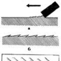
Scheme 12. Classification of blunt objects according to the nature of their traumatic surface.
pom impact is said in cases where mechanical damage is caused by the surface of an object.
The shape and size, mass, strength and elasticity, and the nature of the surface of blunt objects are very diverse. Their kinetic energy at the moment of impact, the location and direction of the acting force are different. All this determines the significant morphological diversity of damage caused by blunt objects. The properties of damage to a certain extent depend on the anatomical and physiological properties of the affected part of the body, the presence of concomitant pathologies and injuries, the age of the victim, the duration of the injury, the nature of healing, etc.
The nature of the damage at the site of application of force is mainly determined by the properties of the traumatic surface of a blunt object. Its main characteristics - size, shape and relief - form the basis for the classification of blunt objects (Diagram 12).
By size, limited and unlimited (wide) traumatic surfaces are distinguished. A surface is considered limited if its boundaries (all or some of them) do not extend beyond the surface of the damaged part of the body. This concept is relative, since the same surface of the same object, depending on the size and shape of the surface of the damaged part of the body, in some cases will be limited, in others it will be unlimited (wide). If the dimensions of the traumatic surface of a blunt object extend beyond the impact area, then such a surface is considered as unlimited. Thus, when hit on the back of the head with the plane of a wide board, the traumatic surface will be unlimited in relation to the surface of the damaged area of the head. If the same plane of the same board strikes the back, then the traumatic surface will be limited in relation to the area of the back. When struck by an object with a limited traumatic surface, the shape and size of the damage are determined primarily by the size and shape of the traumatic surface. When exposed to an object with an unlimited surface, the shape and size of the damage will mainly be determined by the properties of the damaged part of the body.
If it is established that the damage occurred from the action of an object with a limited striking surface, it is imperative to speak out about both the specific shape and the specific dimensions of the traumatic surface of this object.
The shape of the traumatic surface can be flat (triangular, square, rectangular, oval, round, etc.), angular (in the form of a dihedral angle - ribbed or in the form of a polyhedral angle or vertex), curved (spherical, cylindrical, etc.) and combined ( combination of flat and curved, combination of flat and angular surfaces, other combinations). Angular objects have edges, edges, and vertices. Edge - a flat surface bounded on all sides. Rib - line of convergence of two faces. Top - the area of convergence of three or more edges and faces.
The relief of traumatic surfaces and ribs can be even (smooth) and uneven (not smooth, rough, with small protrusions and depressions).
There are four main types of blunt force:
impact, compression, stretching, friction.
Hit -a complex short-term process of interaction between a person’s body (or part of the body) and a blunt object, in which the latter exerts an impulse unilateral centripetal effect on the body or part of the body. The impact action can last less than 0.1-0.01 s.
The shorter the impact time, the more energy is transferred to the affected part of the body and the greater the amount of damage. However, with an ultra-short impact time, a paradoxical effect occurs: the volume of damage becomes smaller, since the damage
Only a small part of the energy of the damaging object is transferred to the given part of the body. The latter option in forensic medical practice occurs in exceptional cases. The impact effect is exerted both by a moving object (for example, a thrown stone, protruding parts of a moving car, etc.) and a stationary one (for example, a blow to the head when falling to the ground); massive objects acting with great force can lead to a concussion of the body or part of the human body.
Constriction -This is the process of interaction of a human body or part of the body with two, usually massive, hard, blunt objects, in which both of these objects, acting towards each other, exert a bilateral centripetal effect on the body or part of the body. The compression time is calculated in seconds, and in some cases in minutes. Of two compressive objects, one is always mobile, the other is most often motionless, for example, pressing a person with the body of a car against motionless objects (a wall of a house, a fence, etc.).
Stretching -This is the process of interaction of a human body or part of the body with two solid objects, which, acting in divergent directions, exert a bilateral centrifugal effect on the body or part of the body. The stretching time is tenths of a second, less often - several seconds. Of two objects, one is always mobile, the other is usually motionless. A stationary object fixes a body or part of a body (for example, a machine body), and another object has an eccentric effect (rotating parts of a machine). An eccentric effect is exerted by a fragment of flat or tubular bone that damages the skin. The impact in this case is complex and is a combination of stretching, piercing, and sometimes cutting actions. However, the main action here is also stretching.
Friction -the process of surface interaction between the damaged surface of a body and the damaging surface of a blunt solid object, in which both contacting surfaces are displaced in a tangential or tangential direction relative to each other. The damaged part of the body, the damaging object, or both can be movable.
DAMAGE
The action of blunt objects causes all types of mechanical damage: abrasions, bruises, wounds, fractures, etc. The morphological features of these injuries make it possible to determine:
- signs (properties) of a traumatic blunt object;
- mechanism of damage formation.
The essence (type) of damage is determined by the type of blunt traumatic impact. Typical for impact action will be bruised wounds, fractures, for compression - flattening of a body part, crushing of organs and tissues, for stretching - torn wounds, skin detachment, for friction - extensive abrasions. At the same time, some types of damage may be the result of different exposure options. Thus, bruises occur both from a blow and from compression, abrasions - from both impact and friction, ruptures of internal organs - from impact, compression and stretching.
Abrasion - This is a superficial damage to the skin that does not extend deeper than the papillary layer. The bottom of the abrasion is initially moist, shiny, and located below the level of the surrounding skin. After a few hours, the bottom dries out and gradually begins to fill with a crust, which is necrotic epithelium and the papillary layer of the dermis. By the end of the first day, the crust reaches the level of the surrounding skin, then rises above it. WITH 4 On the 5th day, epithelialization begins along the borders of the abrasion, and the edges of the crust rise. By the 7-9th day, epithelialization ends and the crust falls off, revealing a pink surface that easily gathers into small folds. By the end of the 2nd week, the place where the abrasion was was no different from the surrounding skin. Abrasions caused by blunt objects can be located on any part of the body surface. The number of abrasions is usually equal to the number of traumatic actions. However, abrasions localized on protruding parts within one area of the body or on several conjugate surfaces of the body can also form from a single action of the wide surface of a blunt object. The size of abrasions ranges from pinpoint to several tens and sometimes hundreds of square centimeters. The area of abrasions depends on the area of the surface of a blunt object in contact with the body and on the length of dynamic contact. With such contact, a blunt object forms an abrasion, the initial part of which is the deepest. At the opposite end, whitish patches of exfoliated epidermis may be noticeable. These morphological features make it possible to establish the direction of movement of a blunt object during the formation of abrasions (or the direction of movement of the body in relation to a stationary blunt object).
The shape of abrasions varies and depends on the shape of the traumatic surface of a blunt object and the mechanism of abrasion formation (Fig. 2). During dynamic contact, a
Rice. 2. Intradermal hemorrhages and abrasions from multiple blows with a stick.
a strip-like abrasion, the width of which may reflect one of the sizes of the traumatic surface of a blunt object. Sometimes on the surface of the abrasion there are multiple rectilinear surface scratches parallel to each other, which arise because the traumatic surface of the object was uneven and rough. When struck or compressed, the shape of the abrasion often follows the shape and surface relief of a blunt object. Elements of the material of the traumatic object, or foreign deposits or contaminants present on the surface of a blunt object may be deposited on the surface of the abrasion. The abrasion allows you to determine:
2. duration of injury;
3. shape, relief, dimensions of the traumatic surface and material of a blunt object, foreign deposits on its surface;
4. direction of movement of the traumatic object or body, if the traumatic object is motionless;
5. place of application of force;
6. option and number of traumatic impacts. Bruising called hemorrhage that permeates the subcutaneous fatty tissue. Initially, the bruise has a blue or blue-purple color, determined by the fact that the coloring matter in the blood is in a state of reduced hemoglobin. From the 3-4th day, the bruise acquires a greenish tint (due to bilirubin and verdocromogen), and from the 7-9th day - a yellowish tint (due to bilirubin). After this period, the bruise usually becomes invisible. However, when the skin is dissected, a brownish hemorrhage (due to hemosiderin) can be seen for a long time in the subcutaneous fatty tissue.
If hemorrhages occur only in the skin, they speak of intradermal hemorrhages. They are usually multiple, small in size and round in shape. The accumulation of blood above (or below) the membranes of the brain in the subcutaneous tissue is called hematoma.
Bruising is typical from the action of a blunt hard object and can have a wide variety of localizations. The shape and size of bruises depend on the shape and size of the traumatic surface of a blunt object. Almost always, one blow with a blunt object will result in one bruise. However, with a strong blow from an elongated object, two elongated bruises may appear, located on either side of the striking surface of such an object. The explanation for this phenomenon comes down to the fact that blood vessels are more resistant to compression than to rupture. Therefore, in the impact zone, the vessels are compressed and maintain their integrity, and at the border of this zone they are stretched and torn.
Bruising reflects:
1. fact of injury and blunt nature of the impact;
2. duration of injury;
3. shape, size and relief of the traumatic surface of a blunt hard object;
4. variant and number of traumatic impacts;
5. place of application of force.
Wound - This is damage that extends deeper than the papillary layer of skin. Wounds resulting from the action of blunt hard objects are divided into bruised, lacerated and bruised-lacerated (with deep injuries, clinicians sometimes talk about a muscle or bone wound, a brain wound, etc.).
Bruised woundsarise from a blow, lacerations - from stretching, bruised-lacerations - from a combination of both mechanisms (most often such wounds arise from a blow with a blunt object acting at an angle).
General signs of a bruised wound: uneven, raw, bruised, and often crushed edges of the wound. In its depths there are whitish connective tissue bridges.
Laceration,with the exception of uneven edges, it does not have the listed characteristics.
The expert significance of a laceration is usually limited to determining the type of traumatic impact (sprain). A bruised wound has incomparably greater forensic information value.
Although bruised wounds can form anywhere on the surface of the body, they are most often found where the bone is closest to the surface of the skin, for example on the head. At the edges of the wound, elements of the material of the traumatic object or traces of foreign deposits on its surface may be found.
Objects with an unlimited traumatic surface form bruised wounds surrounded by a wide continuous deposition. The peculiarity of sedimentation is that it is most pronounced in the central sections, and loses its intensity towards the periphery. Its edges are uneven and smoothly blend into intact skin. The wound can have a variety of shapes (straight-line, triradiate, etc.), which are determined by the structure of the underlying bone. In the center of the wound, there is an area of greatest crushing of soft tissue, from which several tears with relatively sharp ends extend to the sides. The bottom of the rupture is represented by wide connective tissue bridges; in the center of the bottom there are crushed soft tissues. Intact hair often hangs over the bottom of the wound.
The nature of bruised wounds resulting from the action of a limited surface of a blunt object largely depends on its shape and size. The general dimensions of such wounds do not extend beyond the traumatic surface of the object. The edge of a blunt object causes rectilinear wounds, square and rectangular traumatic surfaces form 7" or 77-shaped wounds, triangular - angular, round and oval - C-shaped. The edges of such wounds usually have a narrow edge. The bottom of the wounds is deepened , the connective tissue bridges are narrow, represented by individual fibers and are observed mainly in the area of the wound corners.
The walls of wounds resulting from a perpendicular blow are vertical. When struck at an angle, one of the walls of the wound is beveled, the other is undermined.
Blunt objects acting with a spherical or cylindrical surface cause straight wounds with additional tears at the edges. They are surrounded by relatively widespread sedimentation. The edges of such wounds are often crushed.
Wounds caused by blunt objects indicate:
1. on the variant of traumatic impact (impact, compression, stretching, friction);
2. the duration of the injury;
3. on the blunt nature of the impact;
4. on the number of traumatic impacts;
5. on the shape, size of the traumatic surface and material of the blunt object, the nature of foreign deposits on its surface;
6. to the place, direction and strength of the traumatic impact.
Fracture called damage to bone or cartilage. There are fractures that arise from direct contact traumatic action (direct fractures) and from indirect action (indirect fractures, fractures throughout). Direct fractures allow us to judge the properties of the traumatic object, the type and variant of the traumatic impact, indirect fractures - only about the variant of traumatic blunt impact.
Direct fractures are distinguished by the fact that at the point of contact of the traumatic object with the bone, destruction, crushing and mutual layering of bone structures occur. As a result, small defects are observed at the site of application of force due to chipping of the bone substance. Along the edges of the defect, raised flat bone plates are visible, often layered on top of each other and creating the impression of a tiled roof. Indirect fractures do not have these signs. The edges of direct fractures are a coarsely jagged broken line, while indirect ones are a finely jagged one. These signs make it possible to differentiate direct and indirect fractures of any skeletal bones.
Fractures of long bones can be formed from shear, bending, compression, twisting and tearing.
Bone shiftoccurs from a sharp blow with the edge, edge or narrow limited surface of a blunt object. Shear fractures are always straight. They are transverse or oblique-transverse in nature. At the point where the force is applied, a small chip of the compact substance is formed. Thin cracks extend from the edges of the fracture, the free ends of which indicate the place of impact. Sometimes the ends of cracks extending from opposite edges of the fracture join and form a large fragment, most often diamond-shaped, at the site of the impact.
Bone bendleads to a change in mechanical stress in the bones: a tension zone appears on the convex surface of the bend, and a compression zone appears on the curved surface. Since the bone is less resistant to tension, a transverse crack forms on the convex surface of the diaphysis, which spreads to the lateral surfaces, where it bifurcates. The ends of the crack come together on the compression side, forming a large fragment. Bending of a tubular bone can occur with transverse pressure on the diaphysis (for example, when driven by a car wheel), with longitudinal pressure on a bone, as well as with flexion of a bone, one of the epiphyses of which is fixed.
Bone Compressionin the longitudinal direction underlies the formation of impacted fractures. They are localized in the metadiaphyseal region and represent a local compression destruction of the beam structure, which is often combined with fractures that split the diaphysis in the longitudinal direction. Such fractures occur when falling from a great height onto straight legs.
Twisting the bone represents its rotation around the longitudinal axis while simultaneously fixing one of its (bone) ends. In this case, helical fractures occur (often observed in skiers).
Bone avulsion possible only in the area of tendon attachment. The separated portion of the bone mass is usually small. As a rule, such fractures are observed with sudden tension of the tendons in young subjects with incomplete ossification processes.
Fractures of flat bones depend on the size and shape of the traumatic surface of a blunt hard object and the type of its action: impact or compression. A blow to the place where the force is applied causes unilateral direct fractures.
Objects with a limited striking surface acting with little force may cause linear fracture(crack) expanding in the direction of impact. Several radially diverging fractures may also form at the site of application of force. Additional cracks may arise from some of them, which, connecting and intersecting, can form comminuted fractures in a limited area of the cranial vault. With stronger impacts, depressed fractures are formed, corresponding to the size of the traumatic surface and often being a negative reflection of its shape (Fig. 3). Along the edges of such fractures, step-like fragments can form, which gives grounds to call these fractures terrace-shaped. Impacts of great force can cause a complete displacement of a section of bone with the formation of a perforated fracture, reflecting the shape and size of the traumatic surface of the object (Fig. 4).
A small impact caused by the unlimited surface of a blunt hard object can lead to the formation of one or two or three radiating cracks. With impacts of great force, a focus of splintered fractures, limited by an arcuate crack, is formed at the site of its application. From
Rice. 3. Reconstruction of the striking surface of a blunt object according to the nature of the damage: a) depressed fracture; b) cast of the fracture; c) graphic definition of form striking surface of a blunt object; d) corner of a wooden bar, causing the fracture.
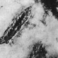
Rice. 4. Perforate fracture of the right parietal bone. On the left is a reinforcing bar that caused a perforated fracture.
Linear cracks diverge radially from this source. The stronger the blow, the larger the area of the focus of comminuted fractures. In the area of focus of comminuted fractures, deformation in the form of flattening of the skull is noticeable.
When compressed, the forces are applied to mutually opposite surfaces of the head and are directed towards each other. In places where force is applied, foci of finely fragmented fractures are formed, surrounded by one or several concentric, arcuate cracks following one after another. The foci of comminuted fractures are united by straight or somewhat curved cracks, showing the direction of compression. Compression is often accompanied by deformation of the head, up to its complete flattening. In rare cases, compression causes a single linear crack to form. It occurs from stretching (cracking) of the bone beyond the point of application of force and is an indirect fracture.
If there are multiple blows to the head, the fracture line created by a subsequent blow will be interrupted by the fracture lines created by previous blows.
When hitting chest at the site of impact, direct, transverse or splintered fractures of the ribs or sternum occur, accompanied by ruptures of the parietal pleura. When compression occurs, multiple bilateral double and triple fractures of the ribs are formed: direct fractures occur at the places where the force is applied, and indirect fractures occur at a distance from the place where the force is applied.
Fractures spine from a local impact lead to splintered fractures of the bodies and processes of individual vertebrae. When forces act along the axis of the spine, compression fractures of the vertebral bodies are formed. With excessively sharp flexion of the spine, dislocations and wedge-shaped compression of the anterior sections of the cervical vertebral bodies most often occur (with extension, the posterior sections). Such fractures are usually accompanied by damage to the ligamentous apparatus of the spine. These fractures are common in traffic accidents, and the mechanism of their occurrence is called whiplash injuries.
When hitting the area pelvis at the site of application of force, unilateral straight single, or double transverse, or comminuted fractures occur. When the pelvis is compressed, bilateral double vertical fractures are formed: direct fractures are found in the places where the force is applied, and indirect fractures of the pelvic bones are found at a distance. Microstructural changes in the fracture zone also make it possible to differentiate the mechanism of damage to the integrity of bone tissue.
Fractures allow us to establish:
1. blunt nature of the impact;
2. fact, type, place, direction, strength and variant of traumatic impact;
3. duration of injury;
4. number and sequence of strikes;
5. shape and size of the traumatic surface of a blunt object.
Damage to internal organs. The morphological features of the internal organs make it possible to make a very limited judgment about the mechanism of action of a blunt solid object and, to an even lesser extent, about its properties. Such injuries are rarely isolated, therefore the mechanism of action and properties of the traumatic object are judged on the basis of an assessment of the morphological signs of the entire set of injuries to soft tissues, bones and internal organs.
Rice. 5. Focal bruises of the cortex of the left frontal and temporal lobes, surrounded by spotty hemorrhages.
When applied to the head, objects of small mass can cause injury only at the site of application of force, where a single injury is observed, including: a bruised wound (less often, an abrasion or bruise), depressed, terraced, comminuted or comminuted-depressed fractures, ruptures of the dura mater and injuries edges of broken bones of brain tissue and soft meninges.
With a head injury, almost any type of intracranial injury and hemorrhage can occur. Of these, the most specific are focal contusions of the cerebral cortex(Fig. 5).
One of the morphological variants of a cortical contusion is the destruction of the cerebral cortex and soft meninges, bordered on the surface by a wide strip of subarachnoid hemorrhages, and in the depths by multiple and small focal hemorrhages with a diameter of less than 1 mm . Such lesions are usually limited by the thickness of the cortex and rarely
occupy the nearest subcortical zone. Another option is characterized by preserving the integrity of the pia mater and the anatomical pattern of the cortex. In this case, the focus of the cortical contusion on the surface of the brain is a group of spotted subarachnoid hemorrhages of round and oval shape with an area of no more than 1 cm2. In the center of the lesion, they can merge with each other, forming a hemorrhage with uneven scalloped edges, surrounded by separate small hemorrhages. A section of this area reveals multiple pinpoint, small-focal or narrow and short strip-like hemorrhages, located mainly in the cortex and in small adjacent areas of the subcortical zone. Foci of contusion involve 1-2 convolutions, less often - the surface and pole of one or two lobes of the brain. The area of cortical bruises is directly proportional to the magnitude of the traumatic force.
The location of the cortical contusions relative to the site of application of force is noteworthy. In impacts from behind, such as falling backwards, they are found at the base and poles of the frontal and temporal lobes. With blows to the head from the front, they are usually localized there and only with blows of extremely high force can they form on the convex surface and poles of the occipital lobes; lateral blows to the head in 2/3 of cases lead to the formation of impact-resistant foci of cortical contusion on the convex surface of the opposite temporal lobe, in 1/3 - cortical contusions occur in the temporal lobe at the site of application of force. In cases where the place of application of force is the parietal region, foci of cortical contusion are found on the basal surface of the frontal and temporal lobes. In these places, bruises of the cortex are found when a force is applied from below, for example, when falling from a great height onto straightened legs and buttocks. Comparison of the place of application of force and the localization of the focus of the cortex bruise in the counter-impact zone makes it possible to determine the direction of the impact.
Focal bruises of the cortex occur during acceleration trauma, when the head comes into contact with an object that significantly exceeds it in mass. As a result of such contact, the movement of the head abruptly accelerates or slows down. This mechanism of injury most often occurs in vehicle injuries and falls from a height. When the head is compressed, focal bruises of the cortex do not occur. The membranes and tissue of the brain can only be injured by bone fragments. The amount of hemorrhage will depend on the diameter of the vessel.
Spinal cord injury occurs only in places where the integrity of the spinal column is damaged in the form of compression fractures and dislocations of the vertebral bodies, ruptures of the ligamentous apparatus and articular capsules. Damage to the brain can range from local intrathecal hemorrhages to complete brain failure. The properties of a traumatic object and the mechanism of its action can be judged mainly by the nature of damage to the bone and ligament-articular structures of the spinal column.
Damage to internal parenchymal organs (liver, kidneys, spleen, etc.) are quite diverse: hemorrhages under their visceral membrane, under the capsule and into the organ tissue, ruptures of the outer membrane, ligamentous apparatus and organ tissue, partial crushing, complete destruction and separation of the organ.
Small superficial hemorrhages and isolated superficial tissue tears are more often formed by strong impacts with objects with a limited traumatic surface. Multiple ruptures of the membranes and tissue of a parenchymal organ, combined with extensive hemorrhages into its tissue, can be the result of either a strong blow from a massive object or compression. Partial crushing or complete destruction most often occurs when a part of the body is compressed by a massive object, for example, when driven over by the wheel of a car or railway transport.
There is no less variety damage to hollow internal organs(stomach, intestines, gall or bladder, etc.): complete or partial ruptures of the organ wall, intrathecal hemorrhages, damage to the ligamentous apparatus and complete separation of the organ. Ruptures of a hollow organ and local hemorrhages in its wall occur from strong impact or compressive action.
Separations of internal parenchymal and hollow organs from their places of attachment, as well as disruption of the integrity of the fixing apparatus of these organs, are observed with strong impacts from massive blunt objects, leading to a general shaking of the body. At the moment of injury, a sharp displacement of the organ occurs, leading to partial or complete rupture of one or more fixing formations (ligaments, arteries, veins, etc.), and with blows of extremely high force - complete separation of the organ.
To morphological characteristics general shock concussion of the body include: hemorrhages in the hilar zone of the lungs, peri-aortic tissue, ligamentous apparatus of the liver and stomach, mesentery of the small intestine, hilum of the kidneys and spleen, ruptures of the hepatic ligaments, gastric ligaments, mesentery of the small intestines, vascular pedicle of the spleen, ureters and renal vessels. The stronger the impact, the greater the volume and severity of the morphological signs of general body concussion.
Morphological features of injuries to internal organs make it possible to establish the fact of injury, type (acceleration injury, compression, etc.), location, direction, number, strength and duration of the traumatic impact.
DAMAGE CAUSED BY HUMAN
In forensic medical practice, injuries caused by nails, fingers, fists, edges of the palm, legs, teeth, and, less commonly, the head, knee, and elbow are often encountered.
With static action, arched abrasions are formed with the free edges of the nails; with dynamic action, stripe-like abrasions are formed.
Pressure with the fingers leads to the appearance of several small round or oval bruises, sometimes combined with arc-shaped or short strip-like abrasions (from nails) located on their background.
Punches or kicks can lead to injuries varying in volume and nature: from superficial abrasions and bruises to bone fractures and ruptures of internal organs. Similar injuries can be caused by the head, elbow, or knee. The volume, nature and location of these damages largely depend on. whether the attacker possessed techniques and skills of special types of wrestling (karate, jiu-jitsu, etc.). However, these damages in no way reflect the properties of the surface relief of the traumatic object.
Rice. 6. Abrasions, bruises and superficial wounds from a bite with human teeth. false sides.
A strike with the edge of the palm can cause significant damage in a limited area. Such blows to the neck can cause dislocations, dislocation fractures, or fractures of the cervical vertebrae, sometimes in combination with damage to the integrity of the spinal cord.
The most typical damage a person can cause is with their teeth (Fig. 6).
Bites cause several abrasions, bruises or superficial wounds. These damages are located in the form of two arched stripes, with their convexities facing in opposite directions.
A steeper arc of damage usually occurs from the action of the teeth of the lower jaw, a flatter one - from the upper jaw. Damage from a bite may also display features of the dental apparatus: malocclusion, gaps in place of missing teeth, atypical structure of one or more teeth, unusual tooth position and DR.
MAIN ISSUES SOLVED DURING FORENSIC MEDICAL EXAMINATION
1. Lifetime and duration of damage.
2. Properties of the traumatic object:
2.1 type of traumatic object;
2.2 traumatic surface (size, shape, relief, overlap);
2.3 mass;
2.4 material;
2.5 the possibility of causing damage with this type of blunt object. 3. Mechanism of damage formation:
3.1 place of application of force;
3.3 type of damaging influence (impact, compression, stretching, friction);
3.4 number of damaging effects;
3.5 impact force;
3.6 relative position of the damaged body part and the damaging object;
3.7 possibility of causing damage under specific conditions.Damage- disruption of the structure and function of organs and tissues at any level: from ultrastructural to the whole organism as a result of the action of physical, chemical, biological and social (mental) environmental factors.
Damage can be uncombined and combined (from the action of two factors).
A blunt object is an object that causes damage with its area or edge.
Injuries from blunt objects are the most common.
The means by which damage can be caused are divided into weapons, instruments and objects.
Weapon- products intended for attack or defense. For example, handguns, finka, dagger, saber, brass knuckles, etc.
Guns - products intended for household or industrial purposes. For example, an axe, table knives, scissors, a hammer, a hoe, a shovel, etc.
Items- all other means that do not have a direct purpose, for example: stone, bottle fragment, brick, etc.
Among mechanical injuries, the impact of blunt hard objects is the most common. This type of injury can occur at home, at work, during transport accidents, in sports and military conditions. Damage can be caused by weapons, tools and objects. Blunt weapons include lead, brass knuckles, nunchucks, flails, etc. Blunt weapons include a hammer, rolling pin, iron, etc. Among the numerous blunt hard objects, damage is most often caused by parts of moving vehicles, various objects that have a certain impact surface area, as well as parts of the human body.
14.3. Mechanisms of formation from the action of blunt hard objects and classification of blunt hard objects
Blunt objects are objects that cause damage by acting mechanically only with their surface . Blunt objects are hard and soft . Violation of the anatomical structure of tissues occurs, as a rule, only when exposed to blunt hard objects . The main mechanisms of damage formation when exposed to blunt hard objects: impact, concussion, compression, stretching, friction.
Hard blunt objects can cause damage to soft tissues, joints, bones, and internal organs.
14.4. Types of mechanical damage from blunt hard objects
Damage to soft tissues - mucous membranes, skin, subcutaneous fat, muscles; damage to joints - ligamentous apparatus, joint capsule; damage to bones - periosteum, bones; internal organs, tears, ruptures, crushing, separation of internal organs. The larger the impact area of the damaging object, the less pronounced the destruction at the point of impact. And the phenomena of body shaking, accompanied by ruptures of internal organs, come to the fore. As the area of the impacting object decreases, more significant damage occurs at the point of impact, since the kinetic energy is concentrated in a small area. The impact is exerted only by the contact part of the object, which, in accordance with its characteristics, causes damage of one form or another. When blunt hard objects act on soft tissues, abrasions, bruises, wounds, and hemorrhages are formed.
 14.4.1. Abrasions
Abrasions - superficial damage to the skin and mucous membranes due to the action of a blunt hard object at an acute angle, with simultaneous sliding and pressure. The action of a blunt solid object is based on impact, compression and friction. A linear abrasion is called a scratch; abrasion is when a significant area of skin is occupied by an abrasion.
Abrasions vary in size, depth, and shape.
During the healing process, the abrasion goes through several stages: first, the abrasion is pinkish-red, shiny, and located below the skin level - on the 1st day, then a crust forms, which is located at the skin level, the crust begins to rise above the skin level on the 2-3rd day; epithelization occurs under the crust (healing process) - 4-6 days; and on days 7-9 the crust disappears. After the crust falls off, a purple area remains; by the end of the second week, the skin color becomes normal. After an abrasion heals, there is never a scar left, since an abrasion is a superficial injury.
Forensic significance of abrasions:
- based on the abrasion, we can talk about the mechanism of damage (abrasions occur from the action of a blunt hard object at an acute angle);
- duration of injury (based on healing of the abrasion);
- direction of action of the traumatic force (deeper abrasion at the beginning, in the end the abrasion is more superficial);
- place of application of force, abrasions are formed at the site of direct impact;
- by the shape of the abrasion one can sometimes talk about the nature of the surface of the object, for example, semilunar abrasions in the neck area are formed from the action of the free edge of the nails when crushed by hands, tread marks in the form of characteristic abrasions; sometimes you can talk specifically about an object if there are inclusions in the abrasion (particles of wood, brick, etc.);
- an abrasion was caused during life or after death; the post-mortem abrasion (parchment stain) is located below the level of intact skin and with a cross-shaped incision in the area of the parchment stain there is no hemorrhage in the underlying tissues.
Bruising
 Bruising are formed by the action of a blunt hard object at a right angle. The action of a blunt hard object is based on impact and compression. Bruises can be superficial, deep, and in size - petechiae, ecchymoses, hematomas.
In the first hours, the bruise is red-purple, red-blue, blue. On days 3-6, the bruise takes on a green tint, and on days 6-10 it turns yellow. Minor bruising disappears after two weeks.
Sometimes it is necessary to differentiate a bruise from a cadaveric spot in the imbibition stage. To distinguish a bruise from a cadaveric spot, it is necessary to make a cross-shaped incision at the site of the bruise; a patch of skin soaked in blood and occupying a limited area is visible.
Forensic significance of bruises:
- we can talk about the mechanism of damage (the action of a blunt solid object at a right angle);
- duration of injury based on change in bruise color;
- the nature of the surface of the object according to the shape of the bruise, for example, the imprint of a belt buckle, tread marks, a bite with teeth, etc.;
- the force of the object;
- the place of application of force, but not always, as for example, with fractures of the bones of the base of the skull, there may be movement of bruises in the area of the eye sockets; when struck in the thigh area, the bruise moves to the popliteal fossa.
 14.4.3. Wounds
Wounds - violation of the entire thickness of the skin and mucous membranes. (see the components of the wound, Fig. 3, page 24, tables and diagrams No. 1.) Wounds are formed as a result of impact, compression, crushing and friction. There are bruised, torn, crushed, scalped, patchwork, bitten, lacerated and bitten. Contusion wounds occur when a direct blow causes tissue to rupture. Crush wounds occur when there is a direct blow with a large amount of crushing. Flap - from an impact made at an angle to the surface of the body, followed by shifting and tearing off the skin in the form of a flap. Scalping wounds most often occur on the head when the skin is torn from the tendon helmet (skull stretching) over a long distance. Lacerations - when the skin is torn. Bite wounds are caused by human teeth. Laceration and bite wounds - from the action of animal teeth.
The most common are bruised wounds, which are often oblong in shape, the edges of the wound are uneven, roughened, bruised, the corners or ends of the wound are rounded (blunt), the depth of the wounds is varied (greater, equal, less than the length of the wound), there are connective tissue bridges in the area of the edges and bottom of the wound , the presence of everted hair follicles in the walls, hemorrhage into the underlying tissue, bone fractures, external bleeding, wound healing is usually poor. After wounds heal, a scar always remains.
 Wounds caused by a blunt hard object in the area of the dorsal surface of the hands, respectively the sternum, ilium, on the front surface of the legs, in the area of the cranial vault usually have smooth edges, sharp ends, a linear shape and often resemble damage from a sharp instrument - cut or chopped wound. The main feature that distinguishes a bruised wound from these wounds is the presence of connective tissue bridges in the area of the edges of the wound, hair follicles in the walls of the wounds are turned out and not cut, a bruised wound is less prone to gaping than a cut one, since around the bruised wound tissues are damaged and lose their contractility.
The shape and size of bruised wounds often, to one degree or another, reflect the characteristics of the striking surface of blunt hard objects.
Forensic significance of wounds:
- place of impact of the weapon,
- mechanism,
- the nature of the traumatic part of the weapon,
- number of traumatic impacts,
- direction of action of the weapon,
- intravitality and postmortemity of the wound,
- how long ago the injury was caused.
Dislocations
Dislocations- displacement of normally contacting articular surfaces and most often occurs in the joints of the upper extremities, less often in the lower ones. This depends on the anatomical structure of the joint and the degree of mobility of the bones in it. Therefore, dislocations especially often occur in the most mobile shoulder and wrist joints.
The skin, as a rule, appears intact, and swelling indicates damage to surrounding tissues (rupture and stretching of the joint capsule, hemorrhage into the joint cavity).
The forensic medical significance of dislocations is that in some cases they make it possible to judge the nature and mechanism of damage. When forensically assessing dislocations, the possibility of habitual and congenital dislocations should be taken into account.
Fractures
Fractures- violation of the anatomical integrity of the bone. Depending on the degree of damage to bone tissue, there are complete and incomplete fractures. Fractures that occur during direct contact with a traumatic object are direct fractures and indirect fractures that arise from an indirect action, for example, during compression.
 In the direction of the fracture line. According to the nature of the fracture, they are distinguished: linear, splintered, multi-splintered, perforated, terrace-shaped. According to communication with the external environment - open and closed.
Forensic medical doctrine of damage is a branch of forensic medicine that studies the patterns of occurrence, variability, research and forensic assessment of damage.
Damage– is a violation of the structure and function of the body as a result of the action of an external damaging factor.
Damaging factor- is an object (blunt and sharp objects, firearms, etc.) or a phenomenon (electricity, high and low temperature, radiant energy, etc.) that has the ability to cause damage (traumatic property). Damaging factors: physical, chemical, biological. Physical are divided into: mechanical, thermal, electrical, barometric and radiation; biological are divided into: microbial and antigenic.
Mechanism of damage formation(mechanism of injury, mechanogenesis of injury) is a complex process of interactions between the damaging factor and the damaged part of the body (or the organism as a whole), which occurs under the influence of the conditional external environment and the properties of the organism itself and leads to the occurrence of damage. Types: impact (fractions of seconds), compression (longer exposure to a blunt object at a right angle), sliding (when an object is applied at an acute angle), stretching, mixed.
Injuries– this is the repetition of homogeneous injuries in people under similar working or living conditions. Types of injuries.
- Transport injuries - combines injuries that occur in people working or using vehicles. There are: ground (wheeled, non-wheeled), underground, air (aviation), water. Wheeled: automobile, motorcycle, bicycle, rail (railway, tram). Non-wheeled: tracked, sled, conveyor, elevator.
- Industrial injuries are a set of injuries that occur in people during the performance of their professional duties. There are: industrial and agricultural injuries.
- Street injuries - combines a group of injuries that occur to people on the street. Street injuries combine mechanical damage associated with falling from a supine position, falling of various objects from a height, conflict situations, etc.
- Household injuries are injuries of very diverse origin that occur in everyday life. Damage that occurs during household work, apartment renovation, use of faulty household appliances, domestic conflicts and other situations.
- Sports injuries - observed in people involved in sports during training or sports competitions.
- Military traumatism is a set of injuries suffered by persons in military service. There are: peacetime military injuries and wartime military injuries - injuries during combat operations (gunshot, explosive, chemical, radiation, thermal, etc.).
With blunt objects in the forensic medical sense, objects that have neither a sharp edge nor a sharp end should be considered.
Based on the area of impact surface, blunt objects are divided into objects with a predominant (wide) and limited traumatic surface; flat or curved (spherical, cylindrical, etc.); smooth or rough; When struck by faceted objects, damage can occur to edges (flat surfaces), edges, and corners.
Based on the nature of the material, blunt objects are divided into hard, soft and crumbly.
The impact of blunt objects causes abrasions, bruises, wounds, dislocations of joints, bone fractures, ruptures and crushing of internal organs, crushing and separation of body parts. By the nature of these injuries, one can judge the mechanism of injury. In some cases, abrasions, bruises and wounds quite clearly reflect the properties of the traumatic object.
Weapon- these are products specifically designed for the purposes of attack and defense (hunting rifle, carbine, saber, brass knuckles, dagger, etc.).
gun- products that are used in everyday life or in production, have a special purpose, but can be used for the purposes of attack or defense (penknife, table knife, straight razor, axe, iron, screwdriver, hammer and similar household items ).
Item- these are any other objects that do not serve any special household or industrial purpose, but can be used for the purpose of attack or defense (stick, brick fragment, stone, glass fragment and others).
It is obvious that determining whether an object is a weapon or an instrument is not within the competence of a forensic expert, but is the prerogative of the judicial investigative authorities. In legal practice, there are cases when a weapon (for example, a penknife, an awl) was recognized as a weapon, since its carrying was intended to attack.
Classification of damage by type:
A. Damage associated with a violation of the anatomical structure:
- Abrasion
- Bruise
- Dislocation
- Fracture
- Gap.
- Kneading
- Dismemberment.
B. Damage associated with impaired physiological function:
- Brain concussion
- Paresis
- Paralysis
- Acoustic trauma
- Accelerotrauma
- Reactive psychoses
- Other functional disorders due to exposure to external factors.
ABRASION- violation of the integrity of the epidermis, which does not penetrate deeper than the papillary layer of the skin. Abrasions are formed when an object acts tangentially, that is, at an angle to the surface of the skin.
Education mechanism abrasions depend on the angle at which the object acted; if it acted at an acute angle, then friction damage occurs at the point of primary contact, and then, when the object penetrates the tissue, pressure is added, in this case the mark of the beginning will be more superficial than the mark graduation. When an object is applied at an angle less than straight, pressure damage occurs at the point of contact with the object, followed by friction damage. In this case, the beginning trace will be deeper than the ending trace. In this case, the abrasion will usually be accompanied by a bruise.
Classification of abrasions according to Solokhin-Bedrin:
- by depth: superficial and deep,
- in shape: straight (scratches), wavy, spindle-shaped, striped, semi-lunar, oval, round, ring-shaped, rectangular, triangular, trapezoidal, diamond-shaped, indefinite.
Forensic medical significance of abrasions:
Abrasions always form directly at the site of traumatic impact (they are an indicator of violence and indicate the location of the application of force). Examination of the edges of abrasions allows one to determine the direction of movement of the traumatic object. In the place where the object first comes into contact with the skin, the edge of the abrasion is even, gentle, sometimes wavy. The opposite edge is usually undermined, steep with preserved, raised, exfoliated scales of the epidermis.
Abrasions allow us to determine how long ago the injury occurred. In the process of regeneration of abrasions, it is customary to distinguish four periods (the time intervals are approximate, since the regeneration of abrasions, as well as other injuries, is influenced by many external and internal factors - the size of the injury, location, age, state of health, presence of diseases, features of the metabolic system, medical assistance, possible re-traumatization in everyday life or work conditions and other conditions.
up to 12 hours - the abrasion has the appearance of a shiny pink wet surface (yellowish or brownish), slightly sunken in comparison with the surrounding intact skin,
12-24 hours - a crust of lymph forms on the surface of the abrasion, and if the surface areas of the papillary layer are damaged, it is mixed with blood.
1-4 days - the crust rises (epithelialization from the periphery to the center), but is not yet rejected.
4-12 days – the edges of the crust are undermined, then the crust peels off from the periphery to the center of the abrasion and completely disappears.
2-3 weeks (up to six months) - depigmentation of the skin, the surface at the site of the fallen off crust is initially pink, but over the course of application this color disappears, the site of the abrasion ceases to differ from the surrounding skin.
According to the observations of V.I. Akopova /1967/ a whitish mark at the site of a former abrasion can sometimes be detected after 30-35 or more days, and by stereomicroscopy - up to several months.
Localization: abrasions on the head and neck enter the last stage within up to 12 days after injury, 14 - 15 days are necessary for epithelization of abrasions on the front surface of the body and up to 20 days on the back and back surface of the lower extremities.
Abrasions make it possible to establish the material from which the traumatic object was made (microscopic particles of the traumatic object can be detected on the surface of the abrasions and in the underlying layers of skin - grains of sand, coal dust, pieces of wood, rust, etc., when carrying out special studies (color print method) it is possible identify areas of metallization and determine the metal from which the traumatic object was made).
The shape and size of abrasions provide information about the shape and size of the object (specific abrasions - crescent-shaped, are formed by strangulation with hands, when the free edges of the fingernails act on the skin. Based on the characteristics of such abrasions (the direction of the convex part, the number of abrasions on the right and left surfaces of the neck), one can determine the relative position of the attacker and the victim, whether the neck was compressed with one or two hands), specific abrasions are formed from the action of teeth and often the characteristics of abrasions reflect the individual characteristics of the structure of the dental apparatus, which makes it possible to subsequently identify the subject who caused the injury; during rape and attempts to rape, abrasions on the inner surfaces of the thighs are typical, abrasions in the shape of parallel or intersecting stripes are characteristic of blows with rods or a whip, abrasions at the openings of the mouth and nose they speak of strangulation or attempts at it, abrasions on the fingers and hands often indicate a struggle and self-defense that preceded death.
Parchment stains– these are postmortem abrasions, they are dense dried areas of skin of yellow or yellow-brown color, if they are located outside the area of cadaveric spots, they differ from intravital abrasions primarily in the absence of crusts (no signs of healing), and there are no hemorrhages during microscopy.
BRUISE- accumulation of blood in the subcutaneous fat, in body cavities or between layers of tissue, resulting from rupture of blood vessels and internal bleeding. There are three fundamentally different groups of bruises: actual bruises in the subcutaneous fat, hematomas (accumulation of blood in body cavities or between layers of tissue), petechiae (point intradermal or intraepithelial hemorrhages caused by ruptures of small vessels).
Actually, bruises are formed when exposed to a blunt traumatic object normal (perpendicular or almost perpendicular) to the surface of the skin. Unlike abrasions, bruises carry less information useful for forensic purposes.
Classification according to Solokhin-Bedrin:
- by origin: traumatic and pathological,
- by place of formation: local and distant (symptom of glasses),
- by time of appearance: early, late, very late,
- by depth: superficial, deep, very deep (subperiosteal),
- by size: petechiae, ecchymoses, large, hematomas,
- in shape: round, oval, rectangular, linear, etc.
Education mechanism: impact, compression, stretching of tissues with blunt objects. When pressure is applied, capillaries rupture; when stretched, larger vessels break (bruises from cupping, Minakov, Vishnevsky, Tardieu spots). As a rule, bruises do not form on the abdomen and buttocks.
Forensic significance:
The location of the bruise does not always correspond to the site of the traumatic impact. Due to the peculiarities of the structure of fatty tissue in some parts of the body, bruises are localized at a distance from the site of injury. Thus, when there is a blow to the area of the glabella or the bridge of the nose, blood flows into the fatty tissue of the eye sockets, simulating the symptom of glasses characteristic of fractures of the base of the skull. Sometimes, when there is a blow to the back surface of the upper third of the thigh, a bruise appears after 1 - 2 days in the popliteal fossa, due to the flow of blood through the interfascial spaces.
The shape and size of the bruise are determined by the volume of blood shed and the architectural features of the fatty tissue at the site of exposure. Typically, bruises are round or oval in shape. Only in rare cases does a bruise reflect the shape of the traumatic object. When struck by objects with an elongated, relatively narrow surface, bruises appear in the form of two parallel stripes, between which there is intact, unpainted skin. This phenomenon is due to the fact that a blow with an elongated narrow object (stick, belt, etc.) is accompanied by squeezing out blood from the vessels at the site of direct impact and rupture of blood vessels at the edges of the active object, where bruises form.
Bruises make it possible to establish the age of origin. In the first hours after formation, the bruise has a purplish-red color due to oxyhemoglobin. Oxyhemoglobin then passes into reduced hemoglobin, the bruise becomes blue-violet with a purple tint. Within 5 - 6 days, the breakdown of blood cells and the subsequent transformation of hemoglobin into methemoglobin and verdochromogen, which is green in color. At this stage, the bruise takes on a greenish tint. Verdochromogen then turns into biliverdinAndbilirubin having a yellow color. At the end of the first and beginning of the second week after the injury, the bruise acquires a yellowish tint. Hemoglobin changes occur unevenly due to varying thickness of the bruise, so the color change occurs from the periphery to the center. After about 7 - 9 days, the bruise becomes three-colored: in the central part - blue-violet, along the periphery - yellow with a brownish tint, and in the intermediate zone - with a pronounced greenish tint. The rate at which a bruise changes color depends on its size, location, age and many other reasons. When analyzing the age of a bruise by changes in its color, it is necessary to take into account that in some areas of the body bruises never bloom. Bruises on the white membranes of the eyes, after the formation of reduced hemoglobin and the acquisition of a blue-violet color, do not undergo further color changes, only gradually discolor, leaving behind areas of gray-yellow coloring that can persist indefinitely. Also, bruises on the transitional border of the lips, on the front surface of the neck, and nail beds do not bloom.
Based on hematomas, one can determine how long ago they formed, as well as the concentration of ethyl alcohol in the blood at the time of the formation of these lesions.
Intravital and postmortem bruises:
- post-mortem bruises (cadaveric spots) are found in all layers of the skin, intravital only in the dermis and pancreas,
- posthumous ones do not bloom,
- post-mortems do not have swelling and compaction of tissues,
- intravital bruising may turn pale when pressed, but does not disappear,
- with a cruciform dissection, there is no blood clot in the post-mortem bruise, and the blood itself is washed off completely with water; in intravital cases, it is not washed off and cannot be removed mechanically.
- Microscopically, postmortems do not have a cellular reaction.
WOUND- this is a violation of the integrity of the skin, penetrating the entire thickness of the skin deeper than the papillary layer, often accompanied by damage to the underlying soft tissues, neurovascular bundles, skeletal bones and internal organs. The totality of damage to the skin and underlying tissues is defined by the concept wound.
Wounds from blunt objects: bruised, crushed, patchwork, scalped, lacerated, bite wounds (so-called bitten).
DISLOCATION- persistent displacement of the articular ends of articulating bones beyond the limits of their physiological mobility (violation of congruence). Depending on the degree of displacement of the articular ends, complete and incomplete (subluxation) dislocations are distinguished. With incomplete dislocation, contact is partially maintained, but in inappropriate places. By origin, it is customary to distinguish traumatic, habitual, congenital and pathological dislocations. Traumatic dislocation occurs as a result of indirect traumatic impact (external force is applied to the peripheral part of the limb) and forced violent movement in the joint. Habitual dislocation is most often the result of improper treatment - traumatic reduction, imperfect or insufficient fixation after reduction. Congenital dislocation is observed in newborns and is associated with abnormal intrauterine development and the formation of defective articular ends. Pathological dislocation is the result of a painful process in the joint cavity or articular ends, for example in osteoarticular tuberculosis, osteomyelitis and other diseases.
In forensic terms, dislocations are injuries that provide little useful information. We can determine the place of application of force (peripheral part of the limb), and very roughly judge the force of impact. It is known that in joints with a large degree of freedom of movement, weak ligamentous apparatus and a small mass of surrounding muscle tissue, dislocations form with relatively small impacts. The greatest force is required to cause a dislocation of the hip joint. Dislocations of the interphalangeal joints of the hand occur quite easily.
FRACTURE– this is a violation of the integrity of bone or cartilage tissue, and is always accompanied by damage to surrounding tissues. According to the mechanism of formation, three groups of fractures are distinguished: direct (local) fractures, that is, damage that occurs at the site of traumatic impact. Secondly, indirect (structural) fractures are formed at a distance from the site of impact and are caused by the deformation of a particular part of the skeleton as a single whole structure. Thirdly, local structural fractures, that is, fractures that begin at the site of impact as direct or local, and then continue as structural (for skull fractures).
According to morphological features, fractures are divided into single and multiple, longitudinal and transverse, oblique and helical, chipped and impacted, depressed, perforated and terrace-shaped, splintered and multi-comminuted, complete and incomplete. Incomplete fractures are sometimes called cracks; they represent a violation of the integrity of the bone, involving only part of the thickness of a particular area (an isolated crack in the internal or external bone plate of the calvarial bones). A special type of fractures is observed in children when the process of ossification of the growth cartilage is not completed; such fractures are called epiphysiolysis (sliding of the epiphyses along the line of the growth cartilage). Fractures can be traumatic and pathological (occur with very minor external influences or even spontaneously in various painful conditions: osteodystrophy, fibrous osteodysplasia, metastases, Paget's disease, osteomyelitis, tuberculosis, etc.).
Forensic significance- fractures remain in a completely skeletonized corpse, and often during the examination of an exhumed corpse, it is the fractures that make it possible to correctly determine the mechanism of damage, the shape features and other group signs of the traumatic object, the severity of the callus during an x-ray examination or during an autopsy provides information to the forensic expert about the possible period that has passed after the fracture occurred, the morphological signs of the fracture itself (its shape, size, condition of the edges and other features) make it possible to establish the direction of action of the external force, the angle at which the force acted on the bone, the shape of the object and its size, force and kinetic energy , spent on the formation of the fracture.
Signs of bone compression:
The fracture line is double, less often single; usually located obliquely, less often transversely; Additional cracks extend from the main fracture line.
The edges of the fracture are uneven, jagged, zigzag, crumpled, with additional cracks; “peaks” often form and the flakes of the compact substance peel off; The edges of the fracture are very poorly aligned with each other, due to chipping of the bone substance (that is, the formation of tiny bone fragments that are lost during examination).
The fracture planes are uneven, coarsely toothed, step-like; the edges of the fracture are usually beveled at an angle of 45° to the surface of the bone, with penetration into each other and compression of the bone substance.
Bone fragments often have a triangle profile and lie freely
Additional cracks extend from the edge of the main fracture line.
In cases of incomplete fractures, deformation of the compact plate in the form of “roller-like swelling”; transverse cracks are noted at the tops of the rollers; often accompanied by detachment of the periosteum and hemorrhages into it.
Signs of bone sprain:
The fracture line is single, usually located transversely, obliquely or spirally.
The edges of the fracture are more or less even; well matched, without signs of chipping; no additional cracks are noted.
The fracture planes are relatively smooth and finely toothed; located vertically relative to the surface of the bone.
There are no bone fragments.
There are no additional cracks.
In cases of incomplete fractures, there is no damage or there are isolated linear cracks.
Breaks– these are closed mechanical damage to soft tissues or internal organs with a violation of their anatomical integrity. There are ruptures of subcutaneous fatty tissue, fascia, muscles, tendons, vessels, nerves, hollow and parenchymal organs. They occur when there is a sufficiently large force of external influence as a result of impact or stretching.
Ruptures of subcutaneous fat are characterized by the formation of extensive hematomas and skin detachment, with the formation of a cavity containing blood clots and crushed fat. Ruptures of the fascia in victims are determined by the presence of a transverse or oblique fissure during palpation examination in a relaxed state, and by its bulging when the muscle is tense. Muscle ruptures in the muscle belly or at the insertion of a tendon occur due to sudden strain or impact to bones (fractures or dislocations). Muscle ruptures are accompanied by hematomas, sharp pain, and dysfunction. In a living person, muscle tears are diagnosed by the presence of a palpable defect, which increases with muscle contraction. At autopsy, the area of the rupture has uneven, blood-soaked edges, a hematoma is pronounced, and a bone fracture or dislocation is determined. Tendon ruptures most often occur when overstretched contracted muscles, less often due to direct traumatic effects, and are localized at the points of attachment to the bone or muscle. A specific sign of tendon rupture is deformation due to the action of antagonist muscles. Ruptures of neurovascular bundles as a result of overstretching or traumatic effects of bone fragments during fractures. Ruptures of hollow and parenchymal organs are always associated with the action of a significant external blunt force and are observed in transport accidents and falls from great heights. At the same time, ruptures of internal organs can also form under local, but concentrated impacts. A punch to the liver area can cause it to rupture. Hollow organs are more susceptible to damage if there is fluid in them; rupture of an overfilled bladder or stomach, distended by food masses, is more likely to occur.
Kneading occur under the action of a significant static load or a load close to static, that is, slowly changing over time. When stretched, the skin, due to its elasticity, has slightly visible damage, while the internal organs, skeletal bones, muscles, and fatty tissue are destroyed. Often, bruises are accompanied by movements of damaged organs or their fragments from one body cavity to another. Similar injuries occur during transport accidents (a body being driven over by the wheels of heavy vehicles), during industrial injuries (collapses in mines) and in some other cases.
Dismemberment body or the separation of individual parts can be observed during direct local impact of blunt and sharp objects (travelling with the wheels of railway transport, the action of chopping or sawing objects), during fixation of the body (or limb) and sudden stretching (casuistic cases of traumatic amputation of a limb have been described when falling from a height), as well as in case of explosive trauma and the deliberate dismemberment of a corpse in order to destroy traces of a crime. The area of dismemberment has specific features that make it possible to determine the mechanism and instrument of injury. Thus, dismemberment when moving by the wheels of a train is fundamentally different from the action of chopping objects, which in turn cannot be confused with the action of sawing or cutting objects. Sometimes the nature of the dismemberment allows a forensic expert to determine the professional affiliation of the person who carried out the dismemberment.
Examination of impact with blunt objects is one of the types of forensic medical examination. Similar studies are carried out to identify the instruments of crime that caused bodily harm to the victim. Examination of exposure to blunt objects is carried out both in relation to living people and in relation to corpses. The objective of the study is to determine the shape and appearance of the object with which the victim was injured. If this is possible, then determine the type of weapon directly.
Examination of impact with blunt objects, like most similar studies, is based on the study of traces left by the crime weapon, that is, on the analysis of existing injuries. Damage is examined, described, classified according to its shape and other characteristics. An idea is formed of the object to which they could have been applied.
A blunt object is a tool and thing of everyday use that affects the body exclusively with its surface. This surface can be smooth or have some textured feature (roughness).
The morphology of bodily injuries caused by blunt objects is extremely diverse, which is explained by their size, shape, elasticity and strength, and surface features. The location and direction of damage and the kinetic energy of the traumatic object also matter.
Speaking about the form of a traumatic object, we can distinguish the following types:
- Flat. At the same time, it can have a regular geometric shape (triangular, oval, square, etc.), a complex shape (for example, star-shaped), and also be of irregular shape.
- Angular. An object has faces, vertices, or edges.
- Curved. Spherical, cylindrical, cone-shaped and other objects have this shape.
- Combined. That is, combining some of the above forms.
Mechanism of injury caused by a blunt object
There are four basic types of blunt force:
- Hit.
- Compression.
- Stretching.
- Friction.
Impact is understood as a short-term process of contact of a blunt object with the body (or part of the body) of a person. In this case, the object exerts a unilateral effect on the human body (part of the body), characterized by an impulsive centripetal nature. The shorter the exposure time, the greater the amount of energy transferred to the victim, thereby causing an increase in the area and volume of injury. Impact can also be caused by a stationary object. The greater the mass of the object and the force applied to it, the more severe the damage.
Compression is characterized by two objects centripetally acting on the body or part of it. When objects are compressed, they move towards each other, and in most cases one of them is movable and the other is not. The compression can be short-term or long-term.
Stretching is the result of the centripetal impact on the body or part of it of two objects moving away from each other. One of the objects is fixed motionless and fixes the body or part of it, and the second moves, moving away from the first.
During friction, an object moves relative to a body while simultaneously coming into contact with it. Both the body and the traumatic object can be mobile.
Types of damage caused by blunt objects
The type of injury depends on how it was inflicted. As a result of impacts, bruised wounds or fractures are formed. Compression causes various types of flattening of body parts, characteristic crushing of tissues and organs. Sprains are characterized by lacerations and skin detachment. For friction - extensive, numerous abrasions occupying a large surface area of the skin. However, it should be understood that the same types of damage can be caused by different exposures. For example, bruising occurs due to both impact and pressure. Abrasions occur from impacts and friction. But ruptures of internal organs can be the result of impact, compression or stretching.
The following types of damage are distinguished:
- Abrasion.
- Bruising (hemorrhage, hematoma).
- Wound.
- Fracture.
- Damage to an internal organ (or several).
- Transport injury.
An abrasion is damage to the upper layers of the skin (not deeper than the papillary layer). Abrasions are formed when the skin comes into tangential contact with blunt objects. If contact occurs with a sharp edge of an object, a linear abrasion is formed, which is more often called a scratch. The number of abrasions in most cases coincides with the number of damaging actions. Exceptions are cases when several protruding parts of the body came into contact with a large surface of the traumatic object. For example, a single fall can cause abrasions on the knees, elbows, palms, etc. The size of the abrasion depends on two parameters: the surface area of the traumatic object and the duration of contact when the blunt object moves along the surface of the body.
During the examination when exposed to blunt objects, analysis of the abrasion allows us to determine:
- force application point;
- properties of the traumatic blunt object;
- direction of traumatic impact;
- duration of damage.
Bruising is an accumulation of blood leaking under pressure from an injured vessel (vessels) in the subcutaneous fatty tissue. At the same time, the integrity of the skin is preserved. Bruising is a characteristic injury caused by blunt objects. They can be localized anywhere in the body. The size of the bruises, as well as their shape, are determined accordingly by the size and shape of the surface of a blunt object in contact with the body. Often the forms of the bruise and the traumatic object coincide. This allows the mechanism of damage to be determined during the examination. For forensic examination, the key point is that when a corpse is struck, no bruising occurs due to the lack of blood circulation in the dead body.
A characteristic property of bruises is a change in color over time. This is due to the chemical transformations of hemoglobin occurring at the site of the bruise. The initial blue-purple color of the bruise changes first to greenish (after 3-4 days), and then to yellow (after 7-9 days).
Hemorrhage is the accumulation of blood leaking from a damaged vessel in the membranes or parenchyma of organs. In some cases, small pinpoint hemorrhages appear in the skin, for example, when a choke loop is applied to the skin of the cervical region.
A hematoma is an accumulation of blood that has leaked from a damaged vessel in a natural or newly formed body cavity. Hematomas can compress vital organs, impairing their functioning.
Analysis of bruises allows you to determine:
- force application point;
- the shape of the traumatic object;
- how long ago the damage was caused.
Wounds include injuries that penetrate deeper than the papillary layer of skin. Characteristic features of wounds include the wound channel and entry hole. Wounds can be blind or through, tangential, penetrating into any body cavity or not, single, combined or multiple. Wounds are also divided into bruised, crushed, lacerated, lacerated and bruised. During the examination, the following is determined:
- properties of the traumatic object;
- direction of movement of the gun;
- the position of the person at the time of injury;
- the possibility (impossibility) of causing a wound independently.
A fracture is a break in the integrity of a bone or cartilage. The fracture can be closed or open. In the latter case, the fracture is accompanied by a wound caused by displacement of a bone fragment. There are also direct and indirect fractures. Direct ones are the result of direct contact with blunt objects, indirect ones are the result of indirect impact, the so-called “fractures along the length”.
Fracture analysis allows us to determine:
- whether there was any violent influence;
- the severity of the damage caused;
- direction of movement of the traumatic object;
- the shape and type of blunt object that caused the fracture.
Damage to internal organs can also result from blunt objects. However, their morphological features provide a very poor idea of the mechanism of application and the properties of the traumatic object. This is due to the distance of the organs from the external boundaries of the body, as a result of which the damage does not have characteristic features indicating signs of a blunt object.
A traffic injury is a bodily injury sustained by a person as a result of contact with a moving vehicle. In most cases, traffic injuries are classified as blunt trauma. The following types of transport injuries are distinguished:
- motorcycle;
- automobile;
- rail;
- aviation.
Fall injuries occur as a result of an impact with an object on which a person falls. A fall can occur from a great height or from one’s own height (so-called flat falls). If nothing prevented the fall, then the injury is the result of a single blow, and then the characteristics of the damage are determined by the relief and size of the surface on which the victim fell. If a body, when falling, hits objects encountered along the way, then such a fall is called a step fall. In this case, the damage is much more complex and numerous.
Damage caused by a person includes injuries caused by blows to parts of the body - fists, ribs of the palms, legs, head, and so on. These injuries also include bites that cause abrasions, bruises, or shallow wounds. It is noteworthy that the lower jaw leaves a steeper arc of marks. Damage caused to the teeth makes it possible to determine the characteristic features of the structure of the jaw and dental apparatus of the attacker: the type of bite, missing teeth, unusual position of the teeth, the special shape of one or more teeth.
In what cases do they resort to examination of impacts with blunt objects?
The examination is carried out during trials related to crimes against health, sexual integrity and human life. The examination is appointed by representatives of the court or investigative authorities, as well as on the initiative of private individuals or their representatives. Most often, examination of exposure to blunt objects is carried out in the following cases:
- The murder weapon must be identified.
- It is necessary to find out what exactly caused the damage.
- It is necessary to establish a correspondence between the item found and the injuries caused.
- It is necessary to determine whether injuries could have been caused by a certain type of object.
- It is necessary to establish the nature and method of injury caused by blunt objects.
What is the legislative framework for the examination of impacts with blunt objects?
Federal Law of May 31, 2001 No. 73-FZ “On State Forensic Expert Activities in the Russian Federation.” Article 25 of the Law describes the procedure for drawing up an expert opinion, as well as the components necessary to be included in it.
What questions should you ask a blunt force trauma examiner?
- What are the characteristics of a blunt object that caused a particular injury?
- What can be said about the instrument based on the characteristics of the wound channel?
- What blunt object could have caused the fracture?
- What is the mechanism of injury?
- What types of injuries did the victim receive?
- Are there any signs that the victim was self-inflicted?
- What is the shape of the object that caused the damage?
- What was the direction of movement of the traumatic object?
- What type of weapon caused the injury?
- What was the position of the victim at the time the wound was inflicted?
- How long has it been since bruising occurred?
- Where was the point of application of force upon impact?
- Are there any signs of abuse?
The list of suggested questions is not exhaustive. If other questions arise, it is advisable to seek advice from an expert before scheduling an examination.
Cost and terms
Forensic examination
A forensic examination is carried out as determined by the court. To appoint an examination to our organization, it is necessary to submit a petition for the appointment of an examination and attach to it an information letter indicating the details of the organization, the possibility of carrying out an examination on the questions raised, the cost and duration of the study, as well as the candidacy of experts indicating their education and work experience. This letter must be certified by the seal of the organization and the signature of its head.
Our specialists prepare an information letter within one working day, after which we send a scanned copy of it by email. Also, if necessary, the original letter can be picked up at the office of our organization. As a rule, the court does not require the original information letter; it is enough to provide a copy of it.
The service for compiling an information letter is provided for free.
Extrajudicial research
Extrajudicial research is carried out on the basis of a contract with 100% prepayment. The agreement can be concluded with both a legal entity and an individual. To conclude an agreement, it is not necessary to be present at the office of our organization; in this case, the sending of all documents, including the expert opinion, will be carried out using the services of postal operators (Dimex, DHL, PonyExpress), which will take no more than 2-4 working days.
Review of expert opinion
A review is necessary in cases where it is necessary to challenge the conclusions of the examination carried out in order to then conduct a repeat study. The conditions for concluding a contract for reviewing are exactly the same as for out-of-court research.
Receiving written expert advice (certificate)
The certificate is not a conclusion, it is of an informational nature and contains answers to questions that do not require a full study, but allow one to assess the feasibility of conducting a full examination.
The conditions for concluding a contract for a certificate are exactly the same as for an out-of-court research.
Obtaining preliminary expert advice
Our specialists are ready to answer any of your questions regarding the conduct of judicial and extrajudicial examinations, assess the feasibility of conducting an examination, provide assistance in formulating research questions, advise on the possibility of conducting a particular analysis, and much more.
Consultation is carried out on the basis of a written request.
To do this, you need to fill out an online application form (or send us a request by e-mail), where you should describe the circumstances of the case in as much detail as possible, formulate the goals that need to be achieved with the help of the examination, preliminary questions, and, if possible, attach all possible documents and descriptions of objects.
The more detailed you tell the circumstances of the case, the more productive the expert’s assistance will be.
Additional services
Reducing the examination period by half
30% to cost
Departure of an expert within the city of Moscow to inspect objects, select samples for research, participate in a court hearing or other events requiring the presence of an expert
Departure of an expert within the Moscow region
Departure of an expert to other regions of Russia
Transport and travel expenses
Preparation of an additional copy of the expert opinion
Legal advice on issues not related to the conduct and appointment of examinations
from 5,000 rub.
Drawing up a statement of claim
Experts
Expert psychiatrist
Coordinator of the International Association of Neuropsychoanalysis in Moscow. Specialist in the field of rehabilitation of people with disabilities due to mental disorders. Engaged in clinical, research, teaching and translation activities.
Dentist, expert
Specialist in the field of aesthetic dentistry and prosthetics. He has original developments on the preservation and restoration of teeth, planning complex interdisciplinary treatment. Takes part in international congresses on aesthetic dentistry, prosthetics and implantology. She trained abroad for a long time with the world's leading doctors in the field of dentistry. Conducts scientific work.
Expert dentist, dentist-therapist of the highest category
Specialist in the field of therapeutic dentistry, orthopedic dentistry, surgical dentistry, healthcare organization and public health, examination of the quality of medical care.
General practitioner, cardiologist, ultrasound diagnostician
Graduated with honors from the Russian National Research Medical University. N.I. Pirogov, specializing in general medicine. Based on the 15th State Clinical Hospital named after. O.M. Filatova completed her residency in therapy. She graduated from a master's degree in economics at Moscow State University, and underwent professional retraining in the specialty of cardiology at the Federal State Budgetary Institution State Scientific Center FMBC named after. A.I. Burnazyan, completed professional training in ultrasound diagnostics at Moscow State Medical University named after. THEM. Sechenov. She worked as a therapist in the 15th City Clinical Hospital named after. O.M. Filatov, senior laboratory assistant and assistant at the Department of Hospital Therapy No. 1 of the Faculty of Medicine of the Russian State Research Medical University named after N.I. Pirogov. She was engaged in teaching and research activities, and took part in clinical studies.
Blunt objects include those that cause damage by acting mechanically only with their surface. Blunt objects can be hard or soft. Direct disruption of the anatomical structure of tissues occurs, as a rule, when exposed to hard, blunt objects. A jet of liquid or gas under high pressure can have a blunt effect. However, such observations are classified as casuistic. Therefore, in what follows, the term blunt object will mean hard, blunt objects. Blunt impact is said to occur when mechanical damage is caused by the surface of an object.
The shape and size, mass, strength and elasticity, and the nature of the surface of blunt objects are very diverse. Their kinetic energy at the moment of impact, the location and direction of the acting force are different. All this determines the significant morphological diversity of damage caused by blunt objects. The properties of damage to a certain extent depend on the anatomical and physiological properties of the affected part of the body, the presence of concomitant pathologies and injuries, the age of the victim, the duration of the injury, the nature of healing, etc.
The nature of the damage at the site of application of force is mainly determined by the properties of the traumatic surface of a blunt object. Its main characteristics - size, shape and relief - form the basis for the classification of blunt objects.
By size, limited and unlimited (wide) traumatic surfaces are distinguished. A surface is considered limited if its boundaries (all or some of them) do not extend beyond the surface of the damaged part of the body. This concept is relative, since the same surface of the same object, depending on the size and shape of the surface of the damaged part of the body, in some cases will be limited, in others it will be unlimited (wide). If the dimensions of the traumatic surface of a blunt object extend beyond the impact area, then such a surface is considered as unlimited. Thus, when hit on the back of the head with the plane of a wide board, the traumatic surface will be unlimited in relation to the surface of the damaged area of the head. If the same plane of the same board strikes the back, then the traumatic surface will be limited in relation to the area of the back. When struck by an object with a limited traumatic surface, the shape and size of the damage are determined primarily by the size and shape of the traumatic surface. When exposed to an object with an unlimited surface, the shape and size of the damage will mainly be determined by the properties of the damaged part of the body.
If it is established that the damage occurred from the action of an object with a limited striking surface, it is imperative to speak out about both the specific shape and the specific dimensions of the traumatic surface of this object.
The shape of the traumatic surface can be flat (triangular, square, rectangular, oval, round, etc.), angular (in the form of a dihedral angle - ribbed or in the form of a polyhedral angle or vertex), curved (spherical, cylindrical, etc.) and combined ( combination of flat and curved, combination of flat and angular surfaces, other combinations). Angular objects have edges, edges, and vertices. Edge - a flat surface bounded on all sides. Rib - line of convergence of two faces. Top - the area of convergence of three or more edges and faces.
The relief of traumatic surfaces and ribs can be even (smooth) and uneven (not smooth, rough, with small protrusions and depressions).
There are four main types of blunt force:
impact, compression, stretching, friction.
Hit - a complex short-term process of interaction between a person’s body (or part of the body) and a blunt object, in which the latter exerts an impulse unilateral centripetal effect on the body or part of the body. The impact action can last less than 0.1-0.01 s.
The shorter the impact time, the more energy is transferred to the affected part of the body and the greater the amount of damage. However, with an ultra-short impact time, a paradoxical effect occurs: the volume of damage becomes smaller, since only a small part of the energy of the damaging object is transferred to the damaged part of the body. The latter option in forensic practice occurs in exceptional cases. The impact effect is exerted both by a moving object (for example, a thrown stone, protruding parts of a moving car, etc.) and a stationary one (for example, a blow to the head when falling to the ground); massive objects acting with great force can lead to a concussion of the body or part of the human body.
Constriction - This is the process of interaction of a human body or part of the body with two, usually massive, hard, blunt objects, in which both of these objects, acting towards each other, exert a bilateral centripetal effect on the body or part of the body. The compression time is calculated in seconds, and in some cases in minutes. Of two compressive objects, one is always mobile, the other is most often motionless, for example, pressing a person with the body of a car against motionless objects (a wall of a house, a fence, etc.).
Stretching - This is the process of interaction of a human body or part of the body with two solid objects, which, acting in divergent directions, exert a bilateral centrifugal effect on the body or part of the body. The stretching time is tenths of a second, less often - several seconds. Of two objects, one is always mobile, the other is usually motionless. A stationary object fixes a body or part of a body (for example, a machine body), and another object has an eccentric effect (rotating parts of a machine). An eccentric effect is exerted by a fragment of flat or tubular bone that damages the skin. The impact in this case is complex and is a combination of stretching, piercing, and sometimes cutting actions. However, the main action here is also stretching.
Friction - the process of surface interaction between the damaged surface of a body and the damaging surface of a blunt solid object, in which both contacting surfaces are displaced in a tangential or tangential direction relative to each other. The damaged part of the body, the damaging object, or both can be movable.
The action of blunt objects causes all types of mechanical damage: abrasions, bruises, wounds, fractures, etc. The morphological features of these injuries make it possible to determine:
Signs (properties) of a traumatic blunt object;
Mechanism of damage formation.
The essence (type) of damage is determined by the type of blunt traumatic impact. Typical for impact action will be bruised wounds, fractures, for compression - flattening of a body part, crushing of organs and tissues, for stretching - lacerations, skin detachment, for friction - extensive abrasions. At the same time, some types of damage may be the result of different exposure options. Thus, bruises occur both from a blow and from compression, abrasions - from both impact and friction, ruptures of internal organs - from impact, compression and stretching.
abrasion - This is a superficial damage to the skin that does not extend deeper than the papillary layer. The bottom of the abrasion is initially moist, shiny, and located below the level of the surrounding skin. After a few hours, the bottom dries out and gradually begins to fill with a crust, which is necrotic epithelium and the papillary layer of the dermis. By the end of the first day, the crust reaches the level of the surrounding skin, then rises above it. WITH 4 On the 5th day, epithelization begins along the borders of the abrasion, and the edges of the crust rise. By the 7-9th day, epithelization ends and the crust falls off, revealing a pink surface that easily gathers into small folds. By the end of the 2nd week, the place where the abrasion was was no different from the surrounding skin. Abrasions caused by blunt objects can be located on any part of the body surface. The number of abrasions is usually equal to the number of traumatic actions. However, abrasions localized on protruding parts within one area of the body or on several conjugate surfaces of the body can also form from a single action of the wide surface of a blunt object. The size of abrasions ranges from pinpoint to several tens and sometimes hundreds of square centimeters. The area of abrasions depends on the area of the surface of a blunt object in contact with the body and on the length of dynamic contact. With such contact, a blunt object forms an abrasion, the initial part of which is the deepest. At the opposite end, whitish patches of exfoliated epidermis may be noticeable. These morphological features make it possible to establish the direction of movement of a blunt object during the formation of abrasions (or the direction of movement of the body in relation to a stationary blunt object).
The shape of abrasions varies and depends on the shape of the traumatic surface of a blunt object and the mechanism of abrasion formation. With dynamic contact, a strip-like abrasion is formed, the width of which may reflect one of the dimensions of the traumatic surface of a blunt object. Sometimes on the surface of the abrasion there are multiple rectilinear surface scratches parallel to each other, which arise because the traumatic surface of the object was uneven and rough. When struck or compressed, the shape of the abrasion often follows the shape and surface relief of a blunt object. Elements of the material of the traumatic object, or foreign deposits or contaminants present on the surface of a blunt object may be deposited on the surface of the abrasion. The abrasion allows you to determine:
2. duration of injury;
3. shape, relief, dimensions of the traumatic surface and material of a blunt object, foreign deposits on its surface;
4. direction of movement of the traumatic object or body, if the traumatic object is motionless;
5. place of application of force;
6. option and number of traumatic impacts.
Bruising called hemorrhage that permeates the subcutaneous fatty tissue. Initially, the bruise has a blue or blue-purple color, determined by the fact that the coloring matter in the blood is in a state of reduced hemoglobin. From the 3-4th day, the bruise acquires a greenish tint (due to bilirubin and verdocromogen), and from the 7-9th day - a yellowish tint (due to bilirubin). After this period, the bruise usually becomes invisible. However, when the skin is dissected, a brownish hemorrhage (due to hemosiderin) can be seen for a long time in the subcutaneous fatty tissue.
If hemorrhages occur only in the skin, they speak of intradermal hemorrhages. They are usually multiple, small in size and round in shape. The accumulation of blood above (or below) the membranes of the brain in the subcutaneous tissue is called hematoma.
Bruising is typical from the action of a blunt hard object and can have a wide variety of localizations. The shape and size of bruises depend on the shape and size of the traumatic surface of a blunt object. Almost always, one blow with a blunt object will result in one bruise. However, with a strong blow from an elongated object, two elongated bruises may appear, located on either side of the striking surface of such an object. The explanation for this phenomenon comes down to the fact that blood vessels are more resistant to compression than to rupture. Therefore, in the impact zone, the vessels are compressed and maintain their integrity, and at the border of this zone they are stretched and torn.
Bruising reflects:
1. fact of injury and blunt nature of the impact;
2. duration of injury;
3. shape, size and relief of the traumatic surface of a blunt hard object;
4. variant and number of traumatic impacts;
5. place of application of force.
Rana - This is damage that extends deeper than the papillary layer of skin. Wounds resulting from the action of blunt hard objects are divided into bruised, lacerated and bruised-lacerated (with deep injuries, clinicians sometimes talk about a muscle or bone wound, a brain wound, etc.).
Bruised wounds arise from a blow, lacerations - from stretching, bruised-lacerations - from a combination of both mechanisms (most often such wounds arise from a blow with a blunt object acting at an angle).
General signs of a bruised wound: uneven, raw, bruised, and often crushed edges of the wound. In its depths there are whitish connective tissue bridges.
Laceration, with the exception of uneven edges, it does not have the listed characteristics.
The expert significance of a laceration is usually limited to determining the type of traumatic impact (sprain). A bruised wound has incomparably greater forensic information value.
Although bruised wounds can form anywhere on the surface of the body, they are most often found where the bone is closest to the surface of the skin, for example on the head. At the edges of the wound, elements of the material of the traumatic object or traces of foreign deposits on its surface may be found.
Objects with an unlimited traumatic surface form bruised wounds surrounded by a wide continuous deposition. The peculiarity of sedimentation is that it is most pronounced in the central sections, and loses its intensity towards the periphery. Its edges are uneven and smoothly blend into intact skin. The wound can have a variety of shapes (straight-line, triradiate, etc.), which are determined by the structure of the underlying bone. In the center of the wound, there is an area of greatest crushing of soft tissue, from which several tears with relatively sharp ends extend to the sides. The bottom of the rupture is represented by wide connective tissue bridges, in the center of the bottom there are crushed soft tissues. Intact hair often hangs over the bottom of the wound.
The nature of bruised wounds resulting from the action of a limited surface of a blunt object largely depends on its shape and size. The general dimensions of such wounds do not extend beyond the traumatic surface of the object. The edge of a blunt object causes straight wounds, square and rectangular traumatic surfaces form 7" or 77-shaped wounds, triangular - angular, round and oval - C-shaped. The edges of such wounds usually have a narrow edge. The bottom of the wounds is deepened, connective woven bridges are narrow, represented by individual fibers and are observed mainly in the area of wound corners.
The walls of wounds resulting from a perpendicular blow are vertical. When struck at an angle, one of the walls of the wound is beveled, the other is undermined.
Blunt objects acting with a spherical or cylindrical surface cause straight wounds with additional tears at the edges. They are surrounded by relatively wide sedimentation. The edges of such wounds are often crushed.
Wounds caused by blunt objects indicate:
1. on the variant of traumatic impact (impact, compression, stretching, friction);
2. the duration of the injury;
3. on the blunt nature of the impact;
4. on the number of traumatic impacts;
5. on the shape, size of the traumatic surface and material of the blunt object, the nature of foreign deposits on its surface;
6. to the place, direction and strength of the traumatic impact.
Fracture called damage to bone or cartilage. There are fractures that arise from direct contact traumatic action (direct fractures) and from indirect action (indirect fractures, fractures throughout). Direct fractures allow us to judge the properties of the traumatic object, the type and variant of the traumatic impact, indirect fractures - only about the variant of traumatic blunt impact.
Direct fractures are distinguished by the fact that at the point of contact of the traumatic object with the bone, destruction, crushing and mutual layering of bone structures occur. As a result, small defects are observed at the site of application of force due to chipping of the bone substance. Along the edges of the defect, raised flat bone plates are visible, often layered on top of each other and creating the impression of a tiled roof. Indirect fractures do not have these signs. The edges of direct fractures are a coarsely jagged broken line, while indirect ones are a finely jagged one. These signs make it possible to differentiate direct and indirect fractures of any skeletal bones.
Fractures of long bones can be formed from shear, bending, compression, twisting and tearing.
Bone shift occurs from a sharp blow with the edge, edge or narrow limited surface of a blunt object. Shear fractures are always straight. They have the character of transverse or oblique transverse. At the point where the force is applied, a small chip of the compact substance is formed. Thin cracks extend from the edges of the fracture, the free ends of which indicate the place of impact. Sometimes the ends of cracks extending from opposite edges of the fracture join and form a large fragment, most often diamond-shaped, at the site of the impact.
Bone bend leads to a change in mechanical stress in the bones: a tension zone appears on the convex surface of the bend, and a compression zone appears on the curved surface. Since the bone is less resistant to tension, a transverse crack forms on the convex surface of the diaphysis, which spreads to the lateral surfaces, where it bifurcates. The ends of the crack come together on the compression side, forming a large fragment. Bending of a tubular bone can occur with transverse pressure on the diaphysis (for example, when driven by a car wheel), with longitudinal pressure on a bone, as well as with flexion of a bone, one of the epiphyses of which is fixed.
Bone Compression in the longitudinal direction underlies the formation of impacted fractures. They are localized in the metadiaphyseal region and represent a local compression destruction of the beam structure, which is often combined with fractures that split the diaphysis in the longitudinal direction. Such fractures occur when falling from a great height onto straight legs.
Twisting the bone represents its rotation around the longitudinal axis while simultaneously fixing one of its (bone) ends. In this case, helical fractures occur (often observed in skiers).
Bone avulsion possible only in the area of tendon attachment. The separated portion of the bone mass is usually small. As a rule, such fractures are observed with sudden tension of the tendons in young subjects with incomplete ossification processes.
Fractures of flat bones depend on the size and shape of the traumatic surface of a blunt hard object and the type of its action: impact or compression. A blow to the place where the force is applied causes unilateral direct fractures.
Objects with a limited striking surface acting with little force may cause linear fracture(crack) expanding in the direction of impact. Several radially diverging fractures may also form at the site of application of force. Additional cracks may arise from some of them, which, connecting and intersecting, can form comminuted fractures in a limited area of the cranial vault. With stronger impacts, depressed fractures are formed, corresponding to the size of the traumatic surface and often being a negative reflection of its shape. Along the edges of such fractures, step-like fragments can form, which gives grounds to call these fractures terrace-shaped. Impacts of great force can cause a complete shift of a section of bone with the formation of a perforated fracture, reflecting the shape and size of the traumatic surface of the object.
A small impact caused by the unlimited surface of a blunt hard object can lead to the formation of one or two or three radiating cracks. When struck with great force, a focus of comminuted fractures, limited by an arcuate crack, is formed at the site of its application. Linear cracks radiate from this source. The stronger the blow, the larger the area of the focus of comminuted fractures. In the area of focus of comminuted fractures, deformation in the form of flattening of the skull is noticeable.
When compressed, the forces are applied to mutually opposite surfaces of the head and are directed towards each other. In places where force is applied, foci of finely fragmented fractures are formed, surrounded by one or several concentric, arcuate cracks following one after another. The foci of comminuted fractures are united by straight or somewhat curved cracks, showing the direction of compression. Compression is often accompanied by deformation of the head, up to its complete flattening. In rare cases, compression causes a single linear crack to form. It occurs from stretching (cracking) of the bone beyond the point of application of force and is an indirect fracture.
If there are multiple blows to the head, the fracture line created by a subsequent blow will be interrupted by the fracture lines created by previous blows.
When hitting chest at the site of impact, direct, transverse or splintered fractures of the ribs or sternum occur, accompanied by ruptures of the parietal pleura. When compression occurs, multiple bilateral double and triple fractures of the ribs are formed: direct fractures occur at the places where the force is applied, and indirect fractures occur at a distance from the place where the force is applied.
Fractures spine from a local impact lead to splintered fractures of the bodies and processes of individual vertebrae. When forces act along the axis of the spine, compression fractures of the vertebral bodies are formed. With excessively sharp flexion of the spine, dislocations and wedge-shaped compression of the anterior sections of the cervical vertebral bodies most often occur (with extension, the posterior sections). Such fractures are usually accompanied by damage to the ligamentous apparatus of the spine. These fractures are common in traffic accidents, and the mechanism of their occurrence is called whiplash injuries.
When hitting the area pelvis at the site of application of force, unilateral straight single, or double transverse, or comminuted fractures occur. When the pelvis is compressed, bilateral double vertical fractures are formed: direct fractures are found in the places where the force is applied, and indirect fractures of the pelvic bones are found at a distance. Microstructural changes in the fracture zone also make it possible to differentiate the mechanism of damage to the integrity of bone tissue.
Fractures allow us to establish:
1. blunt nature of the impact;
2. fact, type, place, direction, strength and variant of traumatic impact;
3. duration of injury;
4. number and sequence of strikes;
5. shape and size of the traumatic surface of a blunt object.
Damage to internal organs. The morphological features of the internal organs make it possible to make a very limited judgment about the mechanism of action of a blunt solid object and, to an even lesser extent, about its properties. Such injuries are rarely isolated, therefore the mechanism of action and properties of the traumatic object are judged on the basis of an assessment of the morphological signs of the entire set of injuries to soft tissues, bones and internal organs.
When applied to the head, objects of small mass can cause injury only at the site of application of force, where a single injury is observed, including: a bruised wound (less often, an abrasion or bruise), depressed, terraced, comminuted or comminuted-depressed fractures, ruptures of the dura mater and injuries edges of broken bones of brain tissue and soft meninges.
With a head injury, almost any type of intracranial injury and hemorrhage can occur. Of these, the most specific are focal contusions of the cerebral cortex.
One of the morphological variants of a cortical contusion is the destruction of the cerebral cortex and soft meninges, bordered on the surface by a wide strip of subarachnoid hemorrhages, and in the depths by multiple and small focal hemorrhages with a diameter of less than 1 mm. Such lesions are usually limited by the thickness of the cortex and rarely
occupy the nearest subcortical zone. Another option is characterized by preserving the integrity of the pia mater and the anatomical pattern of the cortex. In this case, the focus of the cortical contusion on the surface of the brain is a group of spotted subarachnoid hemorrhages of round and oval shape with an area of no more than 1 cm2. In the center of the lesion, they can merge with each other, forming a hemorrhage with uneven scalloped edges, surrounded by separate small hemorrhages. A section of this area reveals multiple pinpoint, small-focal or narrow and short strip-like hemorrhages, located mainly in the cortex and in small adjacent areas of the subcortical zone. Foci of contusion involve 1-2 convolutions, less often - the surface and pole of one or two lobes of the brain. The area of cortical bruises is directly proportional to the magnitude of the traumatic force.
The location of the cortical contusions relative to the site of application of force is noteworthy. In impacts from behind, such as falling backwards, they are found at the base and poles of the frontal and temporal lobes. With blows to the head from the front, they are usually localized there and only with blows of extremely high force can they form on the convex surface and poles of the occipital lobes; lateral blows to the head in 2/3 of cases lead to the formation of impact-resistant foci of cortical contusion on the convex surface of the opposite temporal lobe, in 1/3 - cortical contusions occur in the temporal lobe at the site of application of force. In cases where the place of application of force is the parietal region, foci of cortical contusion are found on the basal surface of the frontal and temporal lobes. In these places, bruises of the cortex are found when a force is applied from below, for example, when falling from a great height onto straightened legs and buttocks. Comparison of the place of application of force and the localization of the focus of the cortex bruise in the counter-impact zone makes it possible to determine the direction of the impact.
Focal bruises of the cortex occur during acceleration trauma, when the head comes into contact with an object that significantly exceeds it in mass. As a result of such contact, the movement of the head abruptly accelerates or slows down. This mechanism of injury most often occurs in vehicle injuries and falls from a height. When the head is compressed, focal bruises of the cortex do not occur. The membranes and tissue of the brain can only be injured by bone fragments. The amount of hemorrhage will depend on the diameter of the vessel.
Spinal cord injury occurs only in places where the integrity of the spinal column is damaged in the form of compression fractures and dislocations of the vertebral bodies, ruptures of the ligamentous apparatus and articular capsules. Damage to the brain can range from local intrathecal hemorrhages to complete brain failure. The properties of a traumatic object and the mechanism of its action can be judged mainly by the nature of damage to the bone and ligament-articular structures of the spinal column.
Damage to internal parenchymal organs(liver, kidneys, spleen, etc.) are quite diverse: hemorrhages under their visceral membrane, under the capsule and into the organ tissue, ruptures of the outer membrane, ligamentous apparatus and organ tissue, partial crushing, complete destruction and separation of the organ.
Small superficial hemorrhages and isolated superficial tissue tears are more often formed by strong impacts with objects with a limited traumatic surface. Multiple ruptures of the membranes and tissue of a parenchymal organ, combined with extensive hemorrhages into its tissue, can be the result of either a strong blow from a massive object or compression. Partial crushing or complete destruction most often occurs when a part of the body is compressed by a massive object, for example, when driven over by the wheel of a car or railway transport.
There is no less variety damage to hollow internal organs(stomach, intestines, gall or bladder, etc.): complete or partial ruptures of the organ wall, intrathecal hemorrhages, damage to the ligamentous apparatus and complete separation of the organ. Ruptures of a hollow organ and local hemorrhages in its wall occur from strong impact or compressive action.
Separations of internal parenchymal and hollow organs from their places of attachment, as well as disruption of the integrity of the fixing apparatus of these organs, are observed with strong impacts from massive blunt objects, leading to a general shaking of the body. At the moment of injury, a sharp displacement of the organ occurs, leading to partial or complete rupture of one or more fixing formations (ligaments, arteries, veins, etc.), and with blows of extremely high force - complete separation of the organ.
To morphological characteristics general shock concussion of the body include: hemorrhages in the hilar zone of the lungs, peri-aortic tissue, ligamentous apparatus of the liver and stomach, mesentery of the small intestine, hilum of the kidneys and spleen, ruptures of the hepatic ligaments, gastric ligaments, mesentery of the small intestines, vascular pedicle of the spleen, ureters and renal vessels. The stronger the impact, the greater the volume and severity of the morphological signs of general body concussion.
Morphological features of injuries to internal organs make it possible to establish the fact of injury, type (acceleration injury, compression, etc.), location, direction, number, strength and duration of the traumatic impact.
In forensic medical practice, injuries caused by nails, fingers, fists, edges of the palm, legs, teeth, and, less commonly, the head, knee, and elbow are often encountered.
With static action, arched abrasions are formed with the free edges of the nails; with dynamic action, stripe-like abrasions are formed.
Pressure with the fingers leads to the appearance of several small round or oval bruises, sometimes combined with arc-shaped or short strip-like abrasions (from nails) located on their background.
Punches or kicks can lead to injuries varying in volume and nature: from superficial abrasions and bruises to bone fractures and ruptures of internal organs. Similar injuries can be caused by the head, elbow, or knee. The volume, nature and location of these damages largely depend on. whether the attacker possessed techniques and skills of special types of wrestling (karate, jiu-jitsu, etc.). However, these damages in no way reflect the properties of the surface relief of the traumatic object.
A strike with the edge of the palm can cause significant damage in a limited area. Such blows to the neck can cause dislocations, dislocation fractures, or fractures of the cervical vertebrae, sometimes in combination with damage to the integrity of the spinal cord.
Bites cause several abrasions, bruises or superficial wounds. These damages are located in the form of two arched stripes, with their convexities facing in opposite directions.
A steeper arc of damage usually occurs from the action of the teeth of the lower jaw, a flatter one - from the upper jaw. Damage from a bite may also display features of the dental apparatus: malocclusion, gaps in place of missing teeth, atypical structure of one or more teeth, unusual tooth position, etc.
DAMAGE FROM FALLS
The damaging object is the object on the surface of which the body falls. The fall height varies widely: from the height of a person to several tens (falls from windows and roofs of buildings, industrial structures, mountain rocks, etc.) and hundreds of meters (in the latter case we are most often talking about falls from aircraft, in including with an unopened parachute).
At direct(unobstructed) fall, the main damage to the human body occurs from a single impact. The nature of these damages is determined by the size and topography of the impact surface. In the vast majority of cases, this surface is unlimited in size. Much less common are falls on free-standing large objects, the area of impact of which is limited to surfaces adjacent to the edges or corners of this object. The relief of the traumatic surface can be relatively flat (asphalt pavement, concrete slabs, etc.) or uneven due to potholes or small objects located on the surface (individual stones, gravel, crushed stone, etc.). The surface can be hard (asphalt, concrete) or relatively soft (sand, soil, turf, etc.).
At indirect(stepped) fall, the body encounters during its movement any protruding objects with a limited traumatic surface (balconies, canopies, awnings, cornices, electrical wires, ropes, etc.). The shape, size and relief of the traumatic elements of these objects are quite diverse.
Falls in confined spaces (for example, shafts, flights of stairs), as well as falls on uneven inclined surfaces: escalator steps, steep mountain slopes, etc. are usually characterized by a stepped nature.
Often, when any structures or their individual structures collapse, various objects fall along with the human body (the so-called unfree fall), which can cause damage both during movement and after the body falls on the ground. The shape, size, relief and hardness of these objects are also very diverse.
For falling from a height Characterized by the occurrence of multiple lesions, usually forming on different parts of the body.
In a direct free fall, damage is formed that has the following typical set of symptoms (regardless of the place where the force is applied):
Little or no external damage;
Unilateral localization of damage;
The presence of fractures far from the place of application of force (so-called fractures along the length, or distant fractures: impacted fractures of the long tubular bones of the lower extremities, compression fractures of the vertebral bodies, ring-shaped fractures of the base of the skull, etc.);
The predominance of the volume of damage to internal organs over the volume of external organs;
Presence of signs of general body concussion (hemorrhages in the para-aortic tissue, hilar zone of the lungs, ligaments of the liver, hilum of the kidneys and spleen, mesentery of the small intestine, etc.).
With strong impacts on the ground, ruptures of hollow and parenchymal organs can occur. The higher the height of the fall, the more pronounced the signs of general body concussion. In a direct free fall on different parts and surfaces of the body, the following injuries are formed: on the head - comminuted fractures of the skull vault, on the buttocks - comminuted fractures of the ischial bones, on the legs - destruction of the heel bones, on the lateral surface of the body - direct and indirect fractures of the ribs on the side of the fall - on the opposite side, on the back - comminuted fractures of the scapula, spinous processes of the vertebrae and multiple direct fractures of the ribs along the paravertebral and scapular lines, on the anterior surface of the body - oblique transverse or comminuted fractures of the sternum, multiple bilateral fractures of the ribs along the parasternal or midclavicular line, injuries to the facial skeleton , fractures of the kneecaps, impacted fractures of the distal metaphyses of the radial bones.
Equally typical for a direct free fall from a height are fractures that form far from the place where the force is applied:
compression fractures of the vertebral bodies and the body of the sternum occur when falling on the buttocks, the plantar surface of the feet of straightened legs and the head, impacted fractures in the area of the metaphyses of the femur and tibia - when falling on the heels; ring-shaped fractures of the base of the skull - when falling on the buttocks and the plantar surface of the feet of straightened legs.
The place where the force is applied when hitting the ground is related to the trajectory of the fall and depends on the height of the fall, the initial posture of the victim, and also on whether the body was given preliminary acceleration (independent repulsion from the support, an extraneous push above or below the center of gravity of the body, etc. ). Depending on the person's condition (for example, alcohol intoxication, unconsciousness), the fall may be uncoordinated. The totality of damage in coordinated and random falls differs.
Damage caused by stepwise and non-free falls has some distinctive features. If all other signs of damage from a fall from a height are preserved (multiplicity, distant fractures, predominance of internal injuries and signs of general body concussion)
are characterized by versatile localization and can be located not only on adjacent, but also on opposite surfaces of the body.
If in a direct free fall, injuries, despite their multiplicity, are formed from a blunt, predominantly impact impact, then in a step-like and non-free fall, along with damage from a blunt impact, torn, puncture, cut and stab wounds can occur.
To establish the mechanism of the fall in a particular case, they resort to the help of an investigative or expert experiment. To do this, an anatomically balanced mannequin is used that simulates the victim’s body. The dummy is dropped under various static and dynamic reference conditions. The trajectory of the fall is recorded using video or high-speed filming, and the final position of the body is recorded on scale diagrams.
A judgment about a specific type of fall is made on the basis of the entire set of available objective data: the morphology of the injuries, the position of the body after the fall, the situation at the scene of the incident, and the results of the experiment.
When a person falls from his own height, they speak of falling on a plane. In this case, the head mainly suffers. At the site of application of force, abrasions, bruises, bruised wounds, and fractures of the facial or cerebral volumes of the skull usually occur. The fracture lines correspond to the direction of the fall.
At falling on the forehead or back of the head At the base and poles of the frontal and temporal lobes, focal bruises of the cerebral cortex are formed: foci of superficial red softening, surrounded by multiple pinpoint and small-focal hemorrhages, affecting mainly the area of the cortex and the nearest subcortical zone. When falling on the temporal region, focal bruises of the cortex are more often localized on the lateral surface of the opposite temporal lobe and less often - in the cortex of the temporal lobe at the site of application of force.
Fall on the posterolateral surface of the head(left or right half of the occipital region) most often leads to the formation of focal bruises of the cortex at the base and poles of the frontal and temporal lobes of the opposite cerebral hemisphere. In a fall with a preliminary shock to the chest, abdomen, or blows to the head, external injuries are usually formed in the area close to the upper parts of the head (parietal region). Intraventricular and subdural hematomas can occur due to any fall mechanism.